Showing posts with label Conservative and Endodontics. Show all posts

Apicoectomy / Root End Surgery

Apicoectomy / Root End Surgery

What is Apicoectomy?

tooth anatomy

Synonyms of Apicoectomy

  1. Root End Surgery
  2. Periradicular Surgery
  3. Periapical Surgery

Introduction

Apicoectomy or root end resection is a type of Endodontic surgery where root end resection is done and periapical pathologies are removed

Objectives for Apicoectomy

  1. Curettage - Effective curettage of periradicular pathologies eg.Therapy resistant granuloma, cysts and foreign body reaction
  2. Resection - Surgical resection of root apex where apical ramification cannot be eliminated by conventional root canal treatment
  3. Inspection - Inspection of periradicular area to rule out causes of failure, inspection of isthmus and trace accessory canals in nonsurgical endodontic cases

Indications for Apicoectomy

  1. Failure of non-surgical Endodontic Treatment
  2. Failure of non-surgical Endodontic Retreatment
  3. Failure of previous surgery
  4. Intracanal breakage of endodontic instruments
  5. Blocked root canals due to calcifications
  6. Periapical granuloma or cyst
  7. Anatomical problems - Endodontist unable to reach to the Apical Constriction due to non-negotiable or blocked canals, severe root curvatures; which prevents adequate cleaning and shaping of apical 3rd of the root canal
  8. Horizontal apical root fracture
  9. Iatrogenic Errors during root canal therapy - ledging of canals, blockage from debris, separated instruments, overfilling of canals and apical canal transporation

Contraindications for Apicoectomy

  1. Inadequate periodontal support
  2. Active uncontrollable periodontal disease
  3. Poor restorability with post-endodontic restoration
  4. Teeth with poor accesibility
  5. Proximity of lesions to imporatnt anatomical structures - eg. Inferior alveolar nerve, Lingual nerve, Mental foramen and Maxillary sinus
  6. Extensive bone involvement
  7. Systemic Complications - eg. Bleeding disorders, Severe heart disease, Immunocompromised patient

Steps in Apicoectomy

  1. Case Diagnosis
    • Preoperative medical history
    • Oral and radiographic examination
    • Patient counselling
  2. Preoperative surgical notes
    • Premedication: Antibiotics
    • Illumination
    • Examination and Inspection instruments: micromirrors, periodontal probes, endodontic explorer (DG16)
    • Incision, Elevation and Curettage instruments: 15c blade and handle, Molts curette no. 2 - 4, Jacquette and mini jacquette curettte, periosteal soft tissue elevator
    • Retraction instruments
    • Osteotomy and apical root resection instruments: Impact air 45° handpiece, Lindemann burs, micromirrors and microexplorers
    • Instruments for preparing root end: microsurgical ultrasonic instruments, MTA root end filling material
    • Irrigational instruments: Stropko irrigator, microsuction
    • Hemostasis instruments and materials
    • Suturing materials: suture, needle, needle holder, scissor, forceps and tweezer
  3. Instrumentation: Dental operating microscope
  4. Anaesthesia / Hemostasis
    • Local anesthesia
    • Vasoconstrictor eg. Epinephrine
  5. Management of Soft Tissues
    • Retain the interdental papilla, gingival contour and manage the frenal attachment in anterior aesthetic zone
    • Flap design, preparation and types:
      1. Triangular flap - Single vertical releasing incision
      2. Rectangular flap - Two vertical releasing incision
      3. Semilunar flap - Limited access to surgical area and more chances of scar formation
      4. Sulcular or full thickness flap - Requires both horizontal and vertical incision. It is a design of choice for endodontic microsurgeries
      5. Submarginal Scalloped Rectangular flap (Luebke-Ochsenbein Flap) - Ideal for crowned teeth when open crown margins after surgery are an aesthetic concern. 2 Vertical incison and 1 Horizontal scalloped incision away from gingival tissue
    • Flap elevation: Molts curette no. 2 - 4 is suitable for both elevation and curetting with minimum trauma
    • Flap Reflection : Reflect the flap along with the periosteum to minimize the bleeding during surgical procedure
    • Flap retraction: Retractors are used for proper visibility to access surgical
  6. Management of Hard Tissues (Ostectomy)
    • Study the periapical radiograph and OPG to have the idea of anatomical structures involved in Root end surgeries
    • Removal of cortical plate to expose the root end in Periapical surgeries
    • High torque and low speed instrument is preferred with external coolants eg. Normal saline or distilled water
  7. Periradicular curettage
    • Periapical pathosis is curetted with Molts curette no. 2 - 4
    • Jacquette 34 - 35 curette can be used to completely remove the granular tissue or cystic pathosis with true cystic lining
  8. Apical Root end resection
    • Control of hemorrhage
    • Apical 3 mm of the root tip is resected perpendicular to the long axis of the root
    • Root resection is carried out with the help of bur in an impact air 45° handpiece
  9. Root end preparation (Retropreparation)
    • Traditionally slow speed burs were used for retropreparation
    • Ultrasonics retrotips are recommended for root end preparation
    • Ultrasonic retrotips eg. KiS ultrasonic tips used in conjunction of ultrasonic units increases the cutting efficiency, leaving smooth dentin surface which results in better adaptation of filling material, fewer microfractures and less leakage
    • Operative area is dried isolated after thorough irrigation with normal saline or distilled water
  10. Root end filling
    • Root end filling materials: eg. MTA, Intermediate Restorative Material (IRM), SuperEBA, Glass ionomer cement, Diaket, Composite resin, Resin ionomer hybrids
    • MTA is the best material of choice for root end filling
    • After restoring root end is burnished to a concave finish
    • After restorative material sets area is irrigated with normal saline and dried with sterile gauze
    • Confirmatory radiograph is taken before closing the surgical area
  11. Soft tissue repositioning
    • Some hemorrhage is allowed before attempting suturing
    • Repositioning of flap and compression
    • Repositioned flap is kept moist with moist gauze until suturing has begun
  12. Suturing
    • Reverse cutting needles are used
    • Medical grade adhesive such as Cyanoacrylates suggested for closure of surgical wound in endodontics
    • Single interrupted, interrupted loop suture, Vertical mattress suture and single sling suturing techniques used in endodontic surgeries
  13. Postsurgical care
    • Oral cavity is cleaned
    • Moist gauze is kept over surgical area
    • Post-operative instructions and medications prescribed

Complications of Apicoectomy

  1. Post-operative pain
  2. Post-operative bleeding
  3. Swelling
  4. Abscess formation
  5. Sinus tract formation
  6. Increases tooth mobility

Endo-Perio Lesion / Endodontic - Periodontic Lesion

Endo-Perio Lesion / Endodontic - Periodontic Lesion

What are Endo-Perio Lesions?

tooth anatomy and periodontal disease

Introduction

Endodontic - Periodontic Lesions are the simultaneous existence of pulpal problems and inflammatory periodontal disease, which affects the diagnosis and treatment planning and affects the sequence of care to be performed

Classification of Endo-Perio Lesions

A) Simon's Classification

  1. Primary Endodontic Lesion
  2. Primary Endodontic Lesion with Secondary Periodontal Involvement
  3. Primary Periodontal Lesion
  4. Primary Periodontal Lesion with Secondary Endodontic Involvement
  5. True Combined Lesion
  6. Concomitant Endodontic and Periodontal Lesion (Added by Belk and Guttman)

B) Oliet and Pollock's Classification (Based on Treatment Protocol)

  1. Lesions that require Endodontic treatment procedures only
  2. Lesions that require Periodontal treatment procedures only
  3. Lesions that requires Combined Endodontic and Periodontic treatment procedures

Pulpoperiodontal Pathways

pulpoperiodontal pathways diagramatic representation

Pathways of Communication between Pulp and Periodontal tissue

  1. Apical foramen
  2. Dentinal tubules
  3. Lateral canals
  4. Periodontal ligament
  5. Alveolar bone
  6. Palatogingival groove
  7. Neural pathways
  8. Vasculolymphatic drainage pathways
  9. Pathological communications due to fractures and perforations

Predisposing Factors of Endo-Perio Lesion

A) Atypical Anatomical Factors:

  1. Malalignment of tooth leading to food impaction and occlusal trauma
  2. Multirooted tooth - additional or fused roots
  3. Additional root canals
  4. Large lateral or accessory canals in coronal and middle section of roots

B) Trauma: Trauma to a tooth can originate from accidental blow, cavity preparation, restorative procedures, orthodontic treatments, tooth separation, malocclusion and detrimental habits

  1. Crown farcture, root fracture or root displacement resulting in irreversible pulpitis, necrosis or periapical disease
  2. Involvement of pulp and disturbance of the periodontal membrane, with the resultant sinus tract draining through periradicular tissue and exiting through the gingival crevice
  3. Trauma along with gingival inflammation leads to deep periodontal pockets
  4. Trauma in case of multirooted teeth leads to furcation involvement
  5. Due to trauma possible cellular changes seen in the pulp or periodontium leading to internal or external bone resorption associated with root perforation

C) Iatrogenic Factors

  1. Perforation into the furcation of multirooted teeth during root canal treatment
  2. Root perforation during biomechanical preparation
  3. Perforation in apical part of curved root during instrumentation

D) Systemic Factors - Systemic Diseases

Primary Endodontic Lesion

  1. Etiology of Primary Endodontic Lesion
    • Dental Caries
    • Restorative procedures
    • Traumatic injuries
  2. Clinical Features of Primary Endodontic Lesion
    • Pain
    • Tenderness to palpation and percussion
    • Sinus tract if present can be traced to the apex of the involved tooth
    • Abnormal response to vitality testing
  3. Treatment plan - Endodontic therapy

Primary Endodontic Lesion with Secondary Periodontal Involvement

  1. Etiology - Progression of Untreated or chronic primary Endodontic lesion
  2. Clinical Feature
    • Plaque and calculus accumulating in the sulcus leading to pocket formation
    • Lowering of epithelial attachment
  3. Treatment Plan - Endodontic treatment as primary line of therapy followed by Secondary Periodontal therapy

Primary Periodontal Lesion

  1. Etiology - Plaque and Calculus
  2. Clinical Feature
    • Horizontal or Angular bone loss
    • Tooth Mobility
    • Normal response to pulp vitality testing
  3. Treatment Plan - Periodontal therapy

Primary Periodontal Lesion with Secondary Endodontic Involvement

  1. Etiology - Periodontal disease progresses apically and involves pulp through apical foramen and lateral or accessory canal
  2. Clinical Feature
    • Horizontal or Angular bone loss
    • Periodontal pocket formation
    • Tooth Mobility
    • Episodes of acute pulpal pain
  3. Treatment Plan - Primary Endodontic treatment followed by long term Periodontal therapy

True Combined Lesion

  1. Etiology - All endodontic and periodontal causes
  2. Clinical Feature - Chronic lesion with gross pulpal and periodontal destruction
  3. Treatment Plan - Endodontic therapy followed by Radisection or Hemisection

Concomitant Endodontic and Periodontal Lesion

  1. Etiology - Distinct etiological factors of endodontic and periodontal disease which do not influence each other
  2. Clinical Feature- Independent clinical features of both Pulpal and Periodontal Diseases
  3. Treatment Plan - Both Pulpal and Periodontal diseases are treated independently

Cracked Tooth Syndrome

Cracked Tooth Syndrome

What is Cracked Tooth Syndrome?

cracked tooth syndrome

Synonyms for Cracked Tooth Syndrome

  • Cracked Cusp Syndrome
  • Split Teeth Syndrome
  • Incomplete Fracture of Posterior Teeth
  • Greenstick Fracture of Crown

Introduction

A tooth that is incompletely cracked but no part of tooth has broken off.

Pattern of Crack

  1. Centrally located crack may extend to pulp
  2. Peripherally located crack (Cuspal Fracture)

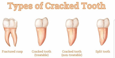
Types of Cracked Tooth Syndrome

  1. Fractured Cusp
  2. Treatable
  3. Non-Treatable
  4. Split Tooth
types of cracked tooth

Clinical Features of Cracked Tooth Syndrome

  1. Pain on biting
  2. Relief after removing pressure
  3. Tenderness positive on Axial Percussion

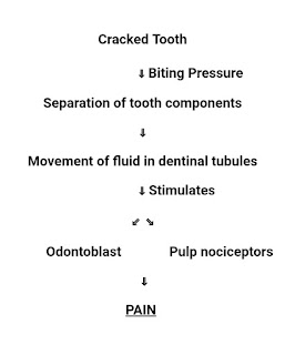
Mechanism of Pain in Cracked Tooth Syndrome

mechanism of pain in cracked tooth syndrome

Diagnosis for Cracked Tooth Syndrome

  1. Clinical Symptoms
  2. Bite Test - Patient is asked to bite upon Tooth Sloth / Toothpick / Cotton Roll
    Inference - When patient bites on tooth sloth it Pains and which gets relieved when biting pressure has withdrawn.

Risk Factor for Cracked Tooth Syndrome

  1. Heavy Restoration
  2. Extensive Dental Restoration
  3. Occlusal Discrepancies (High Points)
  4. Masticatory Accidents

Treatment for Cracked Tooth Syndrome

  1. Small Crack : Remove Compromised part followed by Restoration
  2. Large Crack (No pulp involvement) : Stabilization with Orthodontic Band / Crown / Cast metal Restoration
  3. Large Crack (With pulp involvement) : Stabilization, Endodontic treatment followed by Permanent Restoration
  4. Split Tooth : Extraction (Hopeless Prognosis)