Showing posts with label General Surgery. Show all posts

Raynaud's Disease / Raynaud's Phenomenon

Raynauds's Disease / Raynaud's Phenomenon

What is Raynaud's Disease?

Raynaud's Syndrome

Introduction

Raynaud's Disease is a type of arterial disease also known as Raynaud's Phenomenon and Raynaud's Syndrome

Definition

Raynaud's Disease is a condition characterized by episodic attacks of vasospasm that causes closure of small arteries and arterioles of distal parts of the extremities in response to cold exposure or emotional stimuli

Etiology and Secondary causes of Raynaud's Disease

  1. Cold climate is precipitating factor
  2. Atherosclerosis
  3. Scleroderma
  4. Systemic lupus
  5. Cervical rib
  6. Carpal tunnel syndrome
  7. Vibrating tools
  8. Thoracic outlet syndrome
  9. Cryoglobulinemia
  10. Dermatomyositis

Pathophysiology of Raynaud's Disease

  1. Stage of Syncope: Arterioles undergoes constriction as an abnormal response to cold, hence affected limb becomes blanched and severe pallor develops
  2. Stage of Asphyxia: After a brief period of vasoconstriction, capillaries dilate, filling with deoxygenated blood resulting in bluish discoloration (Cyanosis) of affected part
  3. Stage of Recovery: After vasospasm subsides, vasodilation occurs with reactive hyperemia which produces redness (rubor)

Clinical Features of Raynaud's Disease

  1. Raynaud's Disease affects young females
  2. It causes bilateral episodic digital ischaemia on exposure to cold
  3. Thumb is usually spared
  4. Peripheral pulses are normal
  5. Pallor, Cyanosis and rubor shows colour changes during attack
  6. Sometimes gangrenous patches seen on fingertips due to recurrent attack

Investigation of Raynaud's Disease

  1. Digital Plethysmography
  2. Arteriography
  3. Haemogram
  4. Protein Electrophoresis

Treatment of Raynaud's Disease

A) Conservative Treatment

  • Moving to warm climate
  • Stoppage of Tobacco
  • Avoid unnecessary exposure to cold
  • Occupational exposure should be reduced
  • Calcium antagonist such as Nifedipine

B) Surgical Treatment

  • Cervico-Thoracic Sympathectomy
  • Cervico-Dorsal Sympathectomy
  • Removal of all rami communicating to 2nd or 3rd thoracic ganglion

Varicose Veins of Leg

Varicose Veins of Leg

What are Varicose Veins of Leg?

varicose veins of leg

Introduction

Varicose Veins are the superficial veins of the limbs which becomes dilated, elongated and tortuous

Surgical Anatomy of Varicose Veins of Lower Limb

A) Deep Veins: Deep veins of lower limb shows numerous arteries and branches with there respective valve

  • Posterior tibial veins and its tributaries
  • Peroneal vein
  • Anterior tibial vein
  • Popliteal vein
  • Femoral vein

B) Superficial Veins: Superficial veins lies in the subcutaneous fat between the skin and deep fascia

  • Long (Great) Saphenous vein and its tributaries
  • Short (Small) Saphenous vein and its tributaries

C) Perforating or Communicating Vein: These veins communicate between Superficial and Deep veins, which are of following two types

  • Indirect Perforators
  • Direct Perforators - in the leg, knee and thigh

Etiology of Varicose Veins of Leg

  • Morphological Factor: Erect posture against gravity
  • Congenital cause
  • Prolonged standing: On prolong standing long columns of blood puts pressure on weak valves of veins, which causes failure of veins giving rise to varicosity of long or short saphenous vein
  • Obesity: Excessive fatty tissue in the subcutaneous tissue offers poor support to the veins, leading to formation of varicosity
  • Pregnancy: Progesterone causes dilation and relaxation of veins, which makes valves incompetent, along with that in pregnancy uterus causes pressure on inferior vena cava causing obstruction of venous flow
  • Old age: It causes atrophy / weakness of venous wall and veins gradually becomes incompetent with age
  • Athletes: Forced contraction of calf muscles forces the blood through perforating veins in reverse direction, causing destruction of perforating veins and ulitimately leading to formation of Varicose Veins

Clinical Features of Varicose Veins of Leg

  1. Tired and aching sensation in affected lower limb at the end of day
  2. Sharp pain in grossly dilated veins
  3. Cramps in the calf shortly after retiring to bed
  4. Pain may be bursting or severe in nature particularly localized to the site of incompetent perforating veins
  5. Swollen ankles in evening
  6. Itching and pigmentation over varicosed skin
  7. Eczema of affected skin
  8. Venous ulceration

Local Examination of Varicose Veins of Leg

A) Inspection:

  1. When patient stands up veins becomes prominent
  2. One can feel characteristic thrill when patient coughs
  3. Varix disappears when patient lies down
  4. When the varix is tapped with fingers, a fluid thrill obtained in long saphenous vein
  5. Examination of Venous Ulcer

B) Palpation: Dilated veins are palpated, presence of Cough Impulse is elicited and following tests can be performed

  1. Brodie-Trendelenburg Test - for Sapheno-Femoral valve incompetence
  2. Pratt's Test
  3. Modified Perthe's Test - for deep vein thrombosis
  4. Schwartz's Test - for superficial column of blood
  5. Morrissey's Test / Cough Impulse Test - for Sapheno-Femoral valve incompetence
  6. Fegan's Method - to indicate sites of perforation
  7. Multiple Tourniquet Test - for perforator incompetence

C. Other Examinations includes following:

  1. Abdomen examination - to rule out Pregnancy, Inferior venacaval obstruction eg. Thrombosis
  2. Vaginal or Rectal examination to rule out pelvic tumors
  3. Peripheral arterial pulse examined to exclude presence of arterial insufficiency

Special Investigation of Varicose Veins of Leg

  1. Ascending Phlebography
  2. Thermography
  3. Radioisotope Scanning
  4. Radioactive Fibrinogen Studies
  5. Venography - Ascending and Descending
  6. Doppler Ultrasound
  7. Photoplethysmography
  8. Duplex Ultrasound Imaging

Treatment of Varicose Veins of Leg

  1. Palliative Treatment
    • Avoidance of prolonged standing
    • Creep bandage or elastic stockings are worn throughout the day from toes to thighs, only taken off during sleep
    • Limbs should be raised above the heart
    • Exercise like bicycle riding in the air while lying on the bed, walking etc. should be done to strengthen the calf muscles
  2. Operative Treatment:
    • Indicated for Positive Trendelenburg test and Sapheno-Femoral Incompetence
    • Contraindicated in Pregnancy,Thrombophlebitis and Women taking contraceptive pills
    • Ligation
    • Ligation and Stripping
    • Multiple Cosmetic Phlebectomy
    • Post-Operative Management
    • Fegan's Injection and Compression Treatment

Buerger's Disease / Thromboangiitis Obliterans

Buerger's Disease / Thromboangiitis Obliterans

What is Buerger's Disease ?

buergers disease

Introduction

Buerger's Disease is also known as Thromboangiitis Obliterans which is a type of Inflammatory Disease of Arterial and Venous system. Buerger's Disease was first described by Winiwarter and details were published by Leo Buerger.

Definition of Buerger's Disease

Buerger's Disease is the inflammatory reaction in the arterial wall with the involvement of neighbouring vein and nerve, terminating in thrombosis of the artery

Etiology of Buerger's Disease

  1. Cigarette Smoking: Nicotine produces severe vasospasm and excessive smoking increases the level of carboxyhemoglobin which damage the blood vessels
  2. Lower socioeconomic groups: Recurrent trauma to foot, poor hygiene
  3. Hypercoagulable state
  4. Autonomic hyperactivity
  5. Autoimmune factors
  6. Familial predisposition: Genetic factor
  7. Associated with Rickettsial disease

Pathology of Buerger's Disease

  • Panarteritis: Diffuse inflammatory reaction involving all 3 coats of blood vessels, causing a thrombus which leads to occlusion of lumen (Obliterans)
  • Arteries surrounded by a dense fibrotic reaction which involves neighbouring veins and sometimes nerves
  • Affected superficial vein produces characteristic migratory, recurrent superficial phlebitis
  • Acute Lesion shows acute Arteritis / Periarteritis / Phlebitis / Periphlebitis
  • Chronic Lesion shows arteries and veins bound to each other by Fibrous adhesions

Clinical Features of Buerger's Disease

  1. Thromboangiitis Obliterans predominantly affects male smokers between age group of 20 - 40 years
  2. Foot Claudication: Pain in foot arch while walking due to peripheral involvement of pedal arteries
  3. Intermittent Claudication: Pain is increased when muscle is exercised and disappears when the exercise stops
  4. In chronic ischaemia intermittent claudication progresses to Rest Pain
  5. Gradual postural colour changes
  6. Ulceration and Gangrene of digits and finally the entire foot or hand requires amputation
  7. Phlebitis and ischemic neuritis may also lead to pain
  8. Limbs become rubor or red on dependence and pallor on elevation
  9. Vessels involved are Dorsalis pedis, Posterior tibial and Popliteal

Physical Examination of Buerger's Disease

Inspection:

  • Signs of chronic tissue ischemia are seen before gangrene develops eg. loss of hairs from digits, atrophy of skin, brittle nails
  • Sharply demarcated ischemic area with good circulation to adjacent tissues followed by ulceration and gangrene
  • Gradually gangrene involves whole foot and hand

Palpation:

  • Absence of poserior tibial / dorsalis pedis / radial nerve pulse

Investigations of Buerger's Disease

  1. Patient history
  2. Clinical feature
  3. Arteriography / Angiography: Cock screw / Tree root / Spider leg pattern of vessels, showing the occlusion of small peripheral arteries

Treatment of Buerger's Disease

A) Conservative Treatment

  • Stoppage of Cigarette Smoking
  • Medicines: Vasodilator, Anticoagulants, Dextran, Phenylbutazone and Steroids
  • Prostaglandin therapy prevents platelet aggregation

B) Surgical Treatment

  • Sympathectomy
  • Arterial reconstruction
  • Microvascular transplantation of free omental grafts
  • Amputation in case of severe gangrene

Tracheostomy

Tracheostomy

What is Tracheostomy?

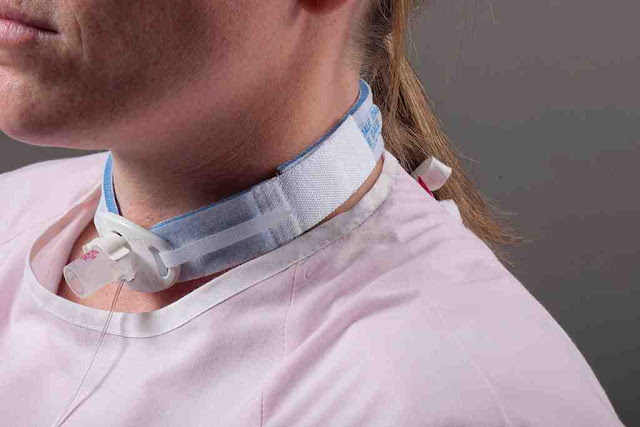
tracheostomy tube

Introduction

Tracheostomy is an operation by which stoma or window is made in the tracheal wall for the purpose of respiration.

Indication of Tracheostomy

  1. Acute and Chronic upper airway obstruction
  2. Maintain patent airway in head and neck surgery
  3. Reduce the risk of pulmonary aspiration
  4. Emergency airway access
  5. Respiratory Paralysis where prolonged intubation is required
  6. Acute Laryngeal Edema eg. Diptheria, Chemical burn and Inhalation of Irritant gases
  7. Excessive tracheobronchial secretion
  8. Foreign body in airway
  9. Injury or pressure to Larynx
  10. Bilateral Abductor Paralysis of vocal cord
  11. Tetanus
  12. Respiratory insufficiency due to Emphysema, Bronchitis, Bronchiectasis

Contraindication of Tracheostomy

  1. Asthma, Chronic Obstructive Pulmonary Disease
  2. Unstable Cervical Spine
  3. Unstable Cardiac Disease
  4. Inability to Cooperate
  5. Severe Hypoxemia

Procedure for Tracheostomy

  1. Preanesthetic medication - Injection atropine 0.6mg half an hour before tracheostomy
  2. Position - The patient is put supine with neck over-extended and chin in the midline
  3. Site - Below isthmus of thyroid gland
  4. Local anesthesia - The part selected is prepared and infiltrated with 2% lignocaine
  5. Procedure - A vertical midline incision is taken about 4 cm long, starting from the suprasternal notch. The skin, platysma and the superficial fascia are cut
  6. The inner margin of sternohyoid muscle is identified and deep layer of cervical fascia is cut
  7. Pretracheal fascia is cut and separated from the trachea
  8. The isthmus is cut between the clamps and its ends ligated
  9. 0.5 ml to 1 ml of lignocaine is injected into the lumen of trachea and fourth and fifth tracheal ring is incised from below upwards
  10. Tracheostomy tube is inserted and tape is tied
  11. The wound of tracheostomy are kept free of secretions by repeated suctions.

Types of Tracheostomy

  1. Emergency
  2. Elective
  3. Permanent

Types of Tracheostomy Tubes

1. Metal Tube : Used for permanent tracheostomy which has inner tube.

metal tracheostomy tube

2. Portex Tube : It has a cuff which can be inflated by injecting the air through outer tube. Balloon gets inflated and secures tracheostomy tube in position.

portex tracheostomy tube

Complications of Tracheostomy

A) During Surgery

  • Injury to trachea and esophagus
  • Injury to recurrent laryngeal nerve
  • False passage

B) Post-operative

  • Blocked tracheostomy tube
  • Surgical emphysema
  • Infection, hemorrhage and ulcerations
  • Pulmonary bronchopneumonia
  • Tracheal stenosis