Showing posts with label Oral Pathology. Show all posts

Osteogenesis Imperfecta

Osteogenesis Imperfecta

What is Osteogenesis Imperfecta?
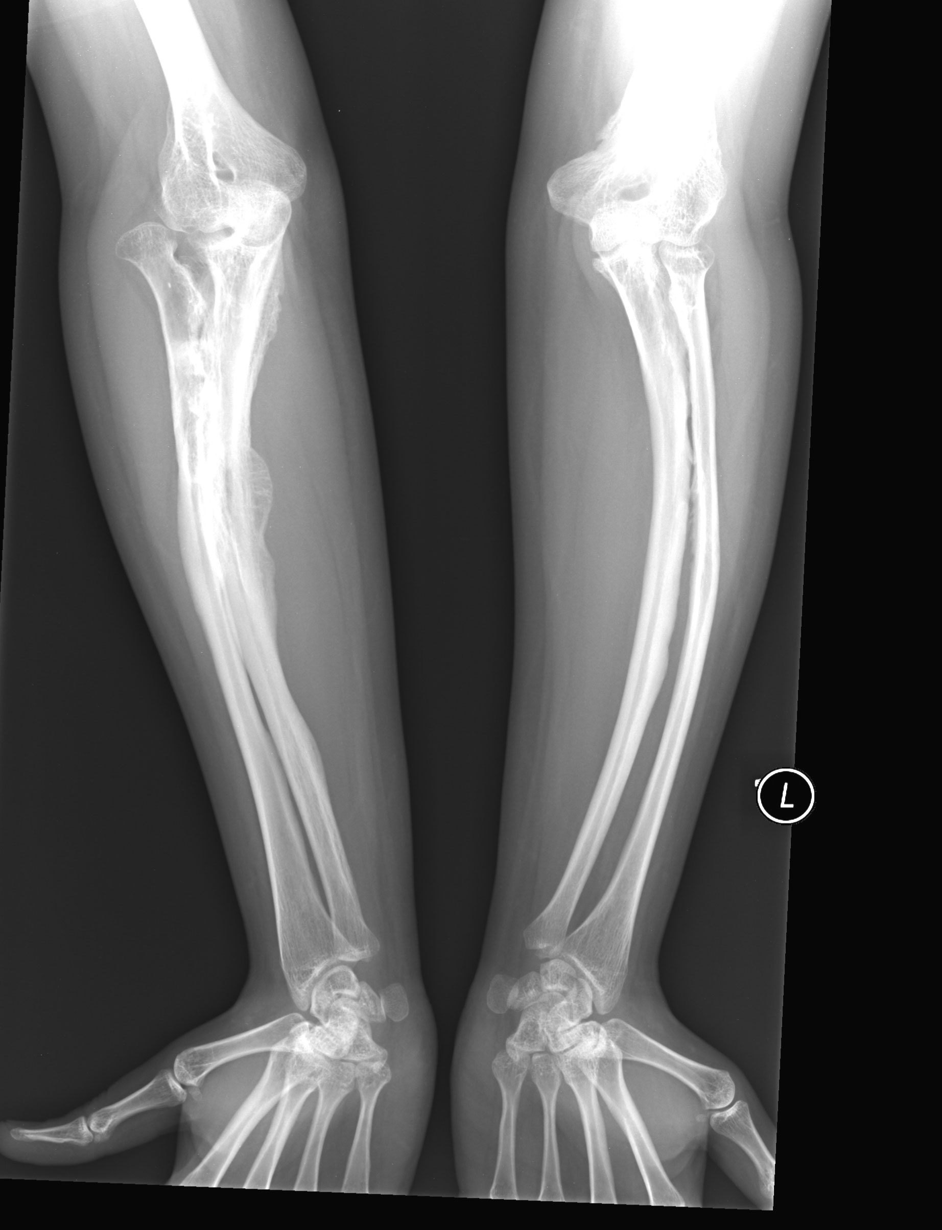
osteogenesis imperfecta type v

Synonyms of Osteogenesis Imperfecta

  • Brittle Bone Disease
  • Fragilitas Ossium
  • Osteopsathyrosis
  • Lobstein’s Disease

Introduction

Osteogenesis Imperfecta is a heterogenous group of disorder with Autosomal Dominant Inheritance Pattern

Etiology of Osteogenesis Imperfecta

etiology of osteogenesis imperfecta

Classification of Osteogenesis Imperfecta

Given by Sillence

Type 1

  • Most common and mildest
  • 10% Prenatal Fracture
  • Blue sclera, Hearing loss
  • Fragile bone, Kyphoscoliosis
  • Sometimes opalscent dentin

Type 2

  • Most severe
  • Extreme bone fragility and fracture
  • 90% Prenatal fracture

Type 3

  • 50% Prenatal fracture
  • All features of type1 except no hearing loss
  • With Opaescent dentin

Type 4

It has 2 subtypes

  • Opalescent dentin is absent
  • Opalescent dentin is present

Clinical Feature of Osteogenesis Imperfecta

  1. Age of onset - Stillborn
  2. Site - Collagenous body part (ex.Bone, Sclera, Fascia, Tendon, Dentin, Skin Fragile and Porous bone) which are more prone to fracture
  3. Fracture heals by Callus formation
  4. Sclera - Pale blue, thin, pigmented choroid giving blue hue
  5. Hearing loss (Hypoacusis) due to compression of auditory nerve by Osteosclerosis
  6. Hypermobility of Joints due to laxity of ligaments
  7. Bleeding capillaries rare
blue sclerae of patient with osteogenesis

Oral Manifestation of Osteogenesis Imperfecta

  1. Abnormal mesodermal calcified tissue
  2. Frontal and Temporal bossing
  3. Abnormal Dentin formation
  4. Tendency of Class 3 malocclusion, Anterior/Posterior Crossbite and Openbite due to Maxillary hypoplasia
  5. Impaction and Ectopic teeth eruption

Radiographic Feature of Osteogenesis Imperfecta

  1. Bowing / Angulate / Deformed fractured bones
  2. Wormian bone in Skull (multiple small sutural bones fail to fuse)
  3. Mixed bony lesions on OPG
  4. Premature pulp obliteration on IOPA

Histopathologic Feature of Osteogenesis Imperfecta

  1. Retarded or abnormal osteoblatic activity
  2. Abnormal Collagen synthesis in organic matrix
  3. Thin cortex
  4. Immature spongy bone
  5. Delicate trabeculae of cancellous bone with microfractures
  6. Defective microvascular system

Treatment of Osteogenesis Imperfecta

  1. Preventive measures
    • Prenatal Ultrasonography
    • Chorion Villus Prenatal Biopsy
  2. Genetic counselling

Malignant Melanoma

Malignant Melanoma

What is Malignant Melanoma?

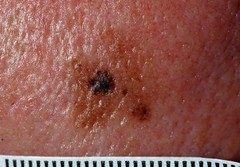
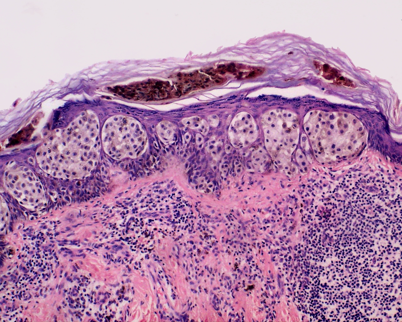
malignant melanoma in situ

Introduction

It is a Malignant Neoplasm of Epidermal Melanocytes and known as most deadliest of Human Neoplasm.

Etiology of Malignant Melanoma

  1. Sunlight Exposure (UV Light)
  2. Artificial UV Source (Tanning Lamp)
  3. Familial - 1st degree relative (2% - 5%)
  4. Xeroderma Pigmentosum
  5. Higher Socioeconomic Status (Outdoor Activities)

Clinicopathologic Classification of Malignant Melanoma

  1. Cutaneous (Most Common)
  2. Non-Cutaneous
  3. Superficial Spreading
  4. Lentigo Maligna
  5. Acral Lentiginous (Most Aggresive)
  6. Amelanotic
  7. Nodular

Clinical Feature of Malignant Melanoma

  1. Incidence - 1/lac/year (Dark Skinned), 50/lac/year (Light Skinned)
  2. Location - Palm, Soles, Mucous Membrane
  3. Precursor Lesion - Acquired Naevus, Dysplastic Naevus, Congenital Naevus, Cellular Blue Naevus

Oral Manifestation Malignant Melanoma

  1. Uncommon in Oral Mucosa (1.6%)
  2. Female Predominance (2×F ˃ M)
  3. Age - 40 - 70 years
  4. Site Predilection - Palate, Maxillary Gingiva, Alveolar Gingiva
  5. Other Sites - Buccal Mucosa, Mandibular Gingiva, Tongue, Lips, Floor of Mouth
  6. Lesion Appearance - Deeply Pigmented, Ulcerated or Hemorrhagic which tends to increase in size
  7. Prodromal Symptoms - Focal Pigmentation (Months or Years back)

Clinical Diagnosis of Malignant Melanoma

( ABCDE Rule )

  • Asymmetry
  • Border - Irregular / Blurred / Notched / Ragged Edges
  • Colour - Pigmentation (Brown / Black /Red / White)
  • Diameter - ˃ 6 mm
  • Elevation - Raised Surface

Histopathologic Feature of Malignant Melanoma

Malignant Melanoma shows Tumor Invasion in deeper histologic cutaneous structure

skin tumors

Clark System (Grades of Tumor Invasion)

Clark Level Characteristics
Level 1 Confined to Epidermis (in situ)
Level 2 Invasion of Papillary Dermis
Level 3 Filling of Papillary Dermis but not extending to Reticular Dermis
Level 4 Invasion of Reticular Dermis
Level 5 Invasion of deep Subcutaneous Tissue

Treatment of Malignant Melanoma

  1. Chemotherapy
  2. Immunotherapy
  3. Surgical - Skin Grafting, Wide Local Incision, Mohs Surgery

Prognosis of Malignant Melanoma

  • Better - Lentigo Maligna (Female Patient)
  • Poor - Nodular, Superficial spreading
  • Worst - Oral Melanotic Lesion