Varicose Veins of Leg

Varicose Veins of Leg

What are Varicose Veins of Leg?

varicose veins of leg

Introduction

Varicose Veins are the superficial veins of the limbs which becomes dilated, elongated and tortuous

Surgical Anatomy of Varicose Veins of Lower Limb

A) Deep Veins: Deep veins of lower limb shows numerous arteries and branches with there respective valve

  • Posterior tibial veins and its tributaries
  • Peroneal vein
  • Anterior tibial vein
  • Popliteal vein
  • Femoral vein

B) Superficial Veins: Superficial veins lies in the subcutaneous fat between the skin and deep fascia

  • Long (Great) Saphenous vein and its tributaries
  • Short (Small) Saphenous vein and its tributaries

C) Perforating or Communicating Vein: These veins communicate between Superficial and Deep veins, which are of following two types

  • Indirect Perforators
  • Direct Perforators - in the leg, knee and thigh

Etiology of Varicose Veins of Leg

  • Morphological Factor: Erect posture against gravity
  • Congenital cause
  • Prolonged standing: On prolong standing long columns of blood puts pressure on weak valves of veins, which causes failure of veins giving rise to varicosity of long or short saphenous vein
  • Obesity: Excessive fatty tissue in the subcutaneous tissue offers poor support to the veins, leading to formation of varicosity
  • Pregnancy: Progesterone causes dilation and relaxation of veins, which makes valves incompetent, along with that in pregnancy uterus causes pressure on inferior vena cava causing obstruction of venous flow
  • Old age: It causes atrophy / weakness of venous wall and veins gradually becomes incompetent with age
  • Athletes: Forced contraction of calf muscles forces the blood through perforating veins in reverse direction, causing destruction of perforating veins and ulitimately leading to formation of Varicose Veins

Clinical Features of Varicose Veins of Leg

  1. Tired and aching sensation in affected lower limb at the end of day
  2. Sharp pain in grossly dilated veins
  3. Cramps in the calf shortly after retiring to bed
  4. Pain may be bursting or severe in nature particularly localized to the site of incompetent perforating veins
  5. Swollen ankles in evening
  6. Itching and pigmentation over varicosed skin
  7. Eczema of affected skin
  8. Venous ulceration

Local Examination of Varicose Veins of Leg

A) Inspection:

  1. When patient stands up veins becomes prominent
  2. One can feel characteristic thrill when patient coughs
  3. Varix disappears when patient lies down
  4. When the varix is tapped with fingers, a fluid thrill obtained in long saphenous vein
  5. Examination of Venous Ulcer

B) Palpation: Dilated veins are palpated, presence of Cough Impulse is elicited and following tests can be performed

  1. Brodie-Trendelenburg Test - for Sapheno-Femoral valve incompetence
  2. Pratt's Test
  3. Modified Perthe's Test - for deep vein thrombosis
  4. Schwartz's Test - for superficial column of blood
  5. Morrissey's Test / Cough Impulse Test - for Sapheno-Femoral valve incompetence
  6. Fegan's Method - to indicate sites of perforation
  7. Multiple Tourniquet Test - for perforator incompetence

C. Other Examinations includes following:

  1. Abdomen examination - to rule out Pregnancy, Inferior venacaval obstruction eg. Thrombosis
  2. Vaginal or Rectal examination to rule out pelvic tumors
  3. Peripheral arterial pulse examined to exclude presence of arterial insufficiency

Special Investigation of Varicose Veins of Leg

  1. Ascending Phlebography
  2. Thermography
  3. Radioisotope Scanning
  4. Radioactive Fibrinogen Studies
  5. Venography - Ascending and Descending
  6. Doppler Ultrasound
  7. Photoplethysmography
  8. Duplex Ultrasound Imaging

Treatment of Varicose Veins of Leg

  1. Palliative Treatment
    • Avoidance of prolonged standing
    • Creep bandage or elastic stockings are worn throughout the day from toes to thighs, only taken off during sleep
    • Limbs should be raised above the heart
    • Exercise like bicycle riding in the air while lying on the bed, walking etc. should be done to strengthen the calf muscles
  2. Operative Treatment:
    • Indicated for Positive Trendelenburg test and Sapheno-Femoral Incompetence
    • Contraindicated in Pregnancy,Thrombophlebitis and Women taking contraceptive pills
    • Ligation
    • Ligation and Stripping
    • Multiple Cosmetic Phlebectomy
    • Post-Operative Management
    • Fegan's Injection and Compression Treatment

Buerger's Disease / Thromboangiitis Obliterans

Buerger's Disease / Thromboangiitis Obliterans

What is Buerger's Disease ?

buergers disease

Introduction

Buerger's Disease is also known as Thromboangiitis Obliterans which is a type of Inflammatory Disease of Arterial and Venous system. Buerger's Disease was first described by Winiwarter and details were published by Leo Buerger.

Definition of Buerger's Disease

Buerger's Disease is the inflammatory reaction in the arterial wall with the involvement of neighbouring vein and nerve, terminating in thrombosis of the artery

Etiology of Buerger's Disease

  1. Cigarette Smoking: Nicotine produces severe vasospasm and excessive smoking increases the level of carboxyhemoglobin which damage the blood vessels
  2. Lower socioeconomic groups: Recurrent trauma to foot, poor hygiene
  3. Hypercoagulable state
  4. Autonomic hyperactivity
  5. Autoimmune factors
  6. Familial predisposition: Genetic factor
  7. Associated with Rickettsial disease

Pathology of Buerger's Disease

  • Panarteritis: Diffuse inflammatory reaction involving all 3 coats of blood vessels, causing a thrombus which leads to occlusion of lumen (Obliterans)
  • Arteries surrounded by a dense fibrotic reaction which involves neighbouring veins and sometimes nerves
  • Affected superficial vein produces characteristic migratory, recurrent superficial phlebitis
  • Acute Lesion shows acute Arteritis / Periarteritis / Phlebitis / Periphlebitis
  • Chronic Lesion shows arteries and veins bound to each other by Fibrous adhesions

Clinical Features of Buerger's Disease

  1. Thromboangiitis Obliterans predominantly affects male smokers between age group of 20 - 40 years
  2. Foot Claudication: Pain in foot arch while walking due to peripheral involvement of pedal arteries
  3. Intermittent Claudication: Pain is increased when muscle is exercised and disappears when the exercise stops
  4. In chronic ischaemia intermittent claudication progresses to Rest Pain
  5. Gradual postural colour changes
  6. Ulceration and Gangrene of digits and finally the entire foot or hand requires amputation
  7. Phlebitis and ischemic neuritis may also lead to pain
  8. Limbs become rubor or red on dependence and pallor on elevation
  9. Vessels involved are Dorsalis pedis, Posterior tibial and Popliteal

Physical Examination of Buerger's Disease

Inspection:

  • Signs of chronic tissue ischemia are seen before gangrene develops eg. loss of hairs from digits, atrophy of skin, brittle nails
  • Sharply demarcated ischemic area with good circulation to adjacent tissues followed by ulceration and gangrene
  • Gradually gangrene involves whole foot and hand

Palpation:

  • Absence of poserior tibial / dorsalis pedis / radial nerve pulse

Investigations of Buerger's Disease

  1. Patient history
  2. Clinical feature
  3. Arteriography / Angiography: Cock screw / Tree root / Spider leg pattern of vessels, showing the occlusion of small peripheral arteries

Treatment of Buerger's Disease

A) Conservative Treatment

  • Stoppage of Cigarette Smoking
  • Medicines: Vasodilator, Anticoagulants, Dextran, Phenylbutazone and Steroids
  • Prostaglandin therapy prevents platelet aggregation

B) Surgical Treatment

  • Sympathectomy
  • Arterial reconstruction
  • Microvascular transplantation of free omental grafts
  • Amputation in case of severe gangrene

Electrocardiogram (ECG) / Electrocardiography

Electrocardiogram (ECG) / Electrocardiography

Basics Of ECG..

electrocardiograph

Introduction

  • Electrocardiogram records the graphical representation of electrical signals from the heart to rule out different heart conditions
  • Electrocardiography is a technique by which electrical activities of the heart is studied
  • Electrocardiograph (ECG Machine) is the instrument by which electrical activities of the heart is recorded
  • Willem Einthoven is called the Father of ECG

Electrocardiographic (ECG) Grid

  1. Duration:
    • Duration of different waves denoted by Vertical lines
    • Interval between 2 thick lines (5 mm) = 0.2 second
    • Interval between 2 thin lines (1 mm) = 0.04 second
  2. Amplitude:
    • Amplitude of ECG wave denoted by Horizontal lines
    • Interval between 2 thick lines (5 mm) = 0.5 mV
    • Interval between 2 thin lines (1 mm) = 0.1 mV
  3. Speed of the Paper: 25 mm per second (normally) or 50 mm per second (high heart rate)

Waves of Electrocardiogram

waves of electrocardiogram
  1. P Wave
    • P wave is the positive wave and 1st wave in ECG
    • It is produced due to the Atrial Depolarization
    • Duration - 0.1 second
    • Amplitude - 0.1 to 0.12 mV
  2. QRS Complex
    • It is also called Initial Ventricular Complex
    • Q wave is small negative wave, continued as tall R wave
    • R wave is positive wave, followed by small negative S wave
    • QRS Complex is produced due to Ventricular Depolarization
    • Duration - 0.08 to 0.10 seconds
    • Amplitude
      • Q wave: 0.1 to 0.2 mV
      • R wave: 1 mV
      • S wave: 0.4 mV
  3. T Wave
    • T wave is the positive wave and final ventricular complex
    • It is produced due to Ventricular Repolarization
    • Duration - 0.2 second
    • Amplitude - 0.3 mV
  4. U Wave
    • It is rare and insignificant wave of ECG
    • It is produced due to Repolarization of papillary muscles

Interval and Segments of ECG

  1. P - R Interval
    • Interval between onset of P wave and onset of Q wave
    • Signifies Atrial Depolarization and conduction of impulses through AV nodes
    • Duration - 0.18 second
  2. Q - T Interval
    • Interval between onset of Q wave and end of T wave
    • Signifies electrical activity in Ventricles
    • Duration - 0.4 to 0.42 seconds
  3. S - T Interval
    • Interval between end of S wave and onset of T wave
    • It is Isoelectric
    • Duration - 0.08 second
  4. R - R Interval
    • Interval between 2 consecutive R waves
    • Signifies the duration of 1 Cardiac CycleInterval between 2 consecutive R waves

ECG Leads

einthoven traingle of ECG
  1. Surface of body is connected to the ECG machine by means of 2 electrodes called ECG lead
  2. ECG leads are of 2 types: Bipolar and Unipolar leads
  3. Einthoven's Triangle: Electrodes are fixed on the limbs (right arm, left arm, left leg) and the heart is in the center of an imaginary equilateral triangle drawn by connecting the roots of these 3 limbs
  4. Bipolar Leads known as standard limb leads where 2 limbs are connected, standard limbs are of 3 following types
    • Lead 1 is obtained by connecting right arm and left arm, where right arm is connected to negative terminal and left arm is connected to positive terminal
    • Lead 2 is obtained by connecting right arm and left leg, where right arm is connected to negative terminal and left leg is connected to positive terminal
    • Lead 3 is obtained by connecting left arm and left leg, where left arm is connected to negative terminal and left leg is connected to positive terminal
  5. Unipolar Leads are of 2 types
    • Unipolar Limb Lead
    • Unipolar Chest Lead

Coleman's Sign

Coleman's Sign

What is Coleman's Sign?

mandibular fracture

Introduction

Coleman's sign is described as Sublingual Hematoma which is a pathognomonic sign of Mandibular body fracture seen as effusion of blood into the tissues of floor of mouth, elevating its mucous membrane and producing a characteristic bluish, tense swelling beneath the tongue

Causes of Coleman's Sign

  1. Road Traffic Accidents
  2. Fall from height
  3. Occupational hazards
  4. Interpersonal fights
  5. Counter Coup fracture

Clinical Features of Coleman's Sign

  1. Mandibular body fracture
  2. Step deformity in chin region
  3. Blood clot seen in floor of mouth (Sublingual Hematoma)
  4. Pain / tenderness
  5. Swelling
  6. Inability to close mouth
  7. Drooling of saliva
  8. Numbness
  9. Soft tissue injury to chin and lower lip
  10. Open bite or mandibular deviation
  11. Obstructive respiration

Treatment Of Coleman's Sign

  1. Proper assessment of case
  2. Radiographic examination of fracture site eg. Panoramic, Lateral Oblique view, posteroanterior view, Occlusal view, Periapical view and CT scan
  3. Soft Tissue management
  4. Analgesics and Antibiotics prescribed
  5. Management of fracture by Reduction, Fixation and Immobilization
  6. Follow up and assessment

Apicoectomy / Root End Surgery

Apicoectomy / Root End Surgery

What is Apicoectomy?

tooth anatomy

Synonyms of Apicoectomy

  1. Root End Surgery
  2. Periradicular Surgery
  3. Periapical Surgery

Introduction

Apicoectomy or root end resection is a type of Endodontic surgery where root end resection is done and periapical pathologies are removed

Objectives for Apicoectomy

  1. Curettage - Effective curettage of periradicular pathologies eg.Therapy resistant granuloma, cysts and foreign body reaction
  2. Resection - Surgical resection of root apex where apical ramification cannot be eliminated by conventional root canal treatment
  3. Inspection - Inspection of periradicular area to rule out causes of failure, inspection of isthmus and trace accessory canals in nonsurgical endodontic cases

Indications for Apicoectomy

  1. Failure of non-surgical Endodontic Treatment
  2. Failure of non-surgical Endodontic Retreatment
  3. Failure of previous surgery
  4. Intracanal breakage of endodontic instruments
  5. Blocked root canals due to calcifications
  6. Periapical granuloma or cyst
  7. Anatomical problems - Endodontist unable to reach to the Apical Constriction due to non-negotiable or blocked canals, severe root curvatures; which prevents adequate cleaning and shaping of apical 3rd of the root canal
  8. Horizontal apical root fracture
  9. Iatrogenic Errors during root canal therapy - ledging of canals, blockage from debris, separated instruments, overfilling of canals and apical canal transporation

Contraindications for Apicoectomy

  1. Inadequate periodontal support
  2. Active uncontrollable periodontal disease
  3. Poor restorability with post-endodontic restoration
  4. Teeth with poor accesibility
  5. Proximity of lesions to imporatnt anatomical structures - eg. Inferior alveolar nerve, Lingual nerve, Mental foramen and Maxillary sinus
  6. Extensive bone involvement
  7. Systemic Complications - eg. Bleeding disorders, Severe heart disease, Immunocompromised patient

Steps in Apicoectomy

  1. Case Diagnosis
    • Preoperative medical history
    • Oral and radiographic examination
    • Patient counselling
  2. Preoperative surgical notes
    • Premedication: Antibiotics
    • Illumination
    • Examination and Inspection instruments: micromirrors, periodontal probes, endodontic explorer (DG16)
    • Incision, Elevation and Curettage instruments: 15c blade and handle, Molts curette no. 2 - 4, Jacquette and mini jacquette curettte, periosteal soft tissue elevator
    • Retraction instruments
    • Osteotomy and apical root resection instruments: Impact air 45° handpiece, Lindemann burs, micromirrors and microexplorers
    • Instruments for preparing root end: microsurgical ultrasonic instruments, MTA root end filling material
    • Irrigational instruments: Stropko irrigator, microsuction
    • Hemostasis instruments and materials
    • Suturing materials: suture, needle, needle holder, scissor, forceps and tweezer
  3. Instrumentation: Dental operating microscope
  4. Anaesthesia / Hemostasis
    • Local anesthesia
    • Vasoconstrictor eg. Epinephrine
  5. Management of Soft Tissues
    • Retain the interdental papilla, gingival contour and manage the frenal attachment in anterior aesthetic zone
    • Flap design, preparation and types:
      1. Triangular flap - Single vertical releasing incision
      2. Rectangular flap - Two vertical releasing incision
      3. Semilunar flap - Limited access to surgical area and more chances of scar formation
      4. Sulcular or full thickness flap - Requires both horizontal and vertical incision. It is a design of choice for endodontic microsurgeries
      5. Submarginal Scalloped Rectangular flap (Luebke-Ochsenbein Flap) - Ideal for crowned teeth when open crown margins after surgery are an aesthetic concern. 2 Vertical incison and 1 Horizontal scalloped incision away from gingival tissue
    • Flap elevation: Molts curette no. 2 - 4 is suitable for both elevation and curetting with minimum trauma
    • Flap Reflection : Reflect the flap along with the periosteum to minimize the bleeding during surgical procedure
    • Flap retraction: Retractors are used for proper visibility to access surgical
  6. Management of Hard Tissues (Ostectomy)
    • Study the periapical radiograph and OPG to have the idea of anatomical structures involved in Root end surgeries
    • Removal of cortical plate to expose the root end in Periapical surgeries
    • High torque and low speed instrument is preferred with external coolants eg. Normal saline or distilled water
  7. Periradicular curettage
    • Periapical pathosis is curetted with Molts curette no. 2 - 4
    • Jacquette 34 - 35 curette can be used to completely remove the granular tissue or cystic pathosis with true cystic lining
  8. Apical Root end resection
    • Control of hemorrhage
    • Apical 3 mm of the root tip is resected perpendicular to the long axis of the root
    • Root resection is carried out with the help of bur in an impact air 45° handpiece
  9. Root end preparation (Retropreparation)
    • Traditionally slow speed burs were used for retropreparation
    • Ultrasonics retrotips are recommended for root end preparation
    • Ultrasonic retrotips eg. KiS ultrasonic tips used in conjunction of ultrasonic units increases the cutting efficiency, leaving smooth dentin surface which results in better adaptation of filling material, fewer microfractures and less leakage
    • Operative area is dried isolated after thorough irrigation with normal saline or distilled water
  10. Root end filling
    • Root end filling materials: eg. MTA, Intermediate Restorative Material (IRM), SuperEBA, Glass ionomer cement, Diaket, Composite resin, Resin ionomer hybrids
    • MTA is the best material of choice for root end filling
    • After restoring root end is burnished to a concave finish
    • After restorative material sets area is irrigated with normal saline and dried with sterile gauze
    • Confirmatory radiograph is taken before closing the surgical area
  11. Soft tissue repositioning
    • Some hemorrhage is allowed before attempting suturing
    • Repositioning of flap and compression
    • Repositioned flap is kept moist with moist gauze until suturing has begun
  12. Suturing
    • Reverse cutting needles are used
    • Medical grade adhesive such as Cyanoacrylates suggested for closure of surgical wound in endodontics
    • Single interrupted, interrupted loop suture, Vertical mattress suture and single sling suturing techniques used in endodontic surgeries
  13. Postsurgical care
    • Oral cavity is cleaned
    • Moist gauze is kept over surgical area
    • Post-operative instructions and medications prescribed

Complications of Apicoectomy

  1. Post-operative pain
  2. Post-operative bleeding
  3. Swelling
  4. Abscess formation
  5. Sinus tract formation
  6. Increases tooth mobility

Endo-Perio Lesion / Endodontic - Periodontic Lesion

Endo-Perio Lesion / Endodontic - Periodontic Lesion

What are Endo-Perio Lesions?

tooth anatomy and periodontal disease

Introduction

Endodontic - Periodontic Lesions are the simultaneous existence of pulpal problems and inflammatory periodontal disease, which affects the diagnosis and treatment planning and affects the sequence of care to be performed

Classification of Endo-Perio Lesions

A) Simon's Classification

  1. Primary Endodontic Lesion
  2. Primary Endodontic Lesion with Secondary Periodontal Involvement
  3. Primary Periodontal Lesion
  4. Primary Periodontal Lesion with Secondary Endodontic Involvement
  5. True Combined Lesion
  6. Concomitant Endodontic and Periodontal Lesion (Added by Belk and Guttman)

B) Oliet and Pollock's Classification (Based on Treatment Protocol)

  1. Lesions that require Endodontic treatment procedures only
  2. Lesions that require Periodontal treatment procedures only
  3. Lesions that requires Combined Endodontic and Periodontic treatment procedures

Pulpoperiodontal Pathways

pulpoperiodontal pathways diagramatic representation

Pathways of Communication between Pulp and Periodontal tissue

  1. Apical foramen
  2. Dentinal tubules
  3. Lateral canals
  4. Periodontal ligament
  5. Alveolar bone
  6. Palatogingival groove
  7. Neural pathways
  8. Vasculolymphatic drainage pathways
  9. Pathological communications due to fractures and perforations

Predisposing Factors of Endo-Perio Lesion

A) Atypical Anatomical Factors:

  1. Malalignment of tooth leading to food impaction and occlusal trauma
  2. Multirooted tooth - additional or fused roots
  3. Additional root canals
  4. Large lateral or accessory canals in coronal and middle section of roots

B) Trauma: Trauma to a tooth can originate from accidental blow, cavity preparation, restorative procedures, orthodontic treatments, tooth separation, malocclusion and detrimental habits

  1. Crown farcture, root fracture or root displacement resulting in irreversible pulpitis, necrosis or periapical disease
  2. Involvement of pulp and disturbance of the periodontal membrane, with the resultant sinus tract draining through periradicular tissue and exiting through the gingival crevice
  3. Trauma along with gingival inflammation leads to deep periodontal pockets
  4. Trauma in case of multirooted teeth leads to furcation involvement
  5. Due to trauma possible cellular changes seen in the pulp or periodontium leading to internal or external bone resorption associated with root perforation

C) Iatrogenic Factors

  1. Perforation into the furcation of multirooted teeth during root canal treatment
  2. Root perforation during biomechanical preparation
  3. Perforation in apical part of curved root during instrumentation

D) Systemic Factors - Systemic Diseases

Primary Endodontic Lesion

  1. Etiology of Primary Endodontic Lesion
    • Dental Caries
    • Restorative procedures
    • Traumatic injuries
  2. Clinical Features of Primary Endodontic Lesion
    • Pain
    • Tenderness to palpation and percussion
    • Sinus tract if present can be traced to the apex of the involved tooth
    • Abnormal response to vitality testing
  3. Treatment plan - Endodontic therapy

Primary Endodontic Lesion with Secondary Periodontal Involvement

  1. Etiology - Progression of Untreated or chronic primary Endodontic lesion
  2. Clinical Feature
    • Plaque and calculus accumulating in the sulcus leading to pocket formation
    • Lowering of epithelial attachment
  3. Treatment Plan - Endodontic treatment as primary line of therapy followed by Secondary Periodontal therapy

Primary Periodontal Lesion

  1. Etiology - Plaque and Calculus
  2. Clinical Feature
    • Horizontal or Angular bone loss
    • Tooth Mobility
    • Normal response to pulp vitality testing
  3. Treatment Plan - Periodontal therapy

Primary Periodontal Lesion with Secondary Endodontic Involvement

  1. Etiology - Periodontal disease progresses apically and involves pulp through apical foramen and lateral or accessory canal
  2. Clinical Feature
    • Horizontal or Angular bone loss
    • Periodontal pocket formation
    • Tooth Mobility
    • Episodes of acute pulpal pain
  3. Treatment Plan - Primary Endodontic treatment followed by long term Periodontal therapy

True Combined Lesion

  1. Etiology - All endodontic and periodontal causes
  2. Clinical Feature - Chronic lesion with gross pulpal and periodontal destruction
  3. Treatment Plan - Endodontic therapy followed by Radisection or Hemisection

Concomitant Endodontic and Periodontal Lesion

  1. Etiology - Distinct etiological factors of endodontic and periodontal disease which do not influence each other
  2. Clinical Feature- Independent clinical features of both Pulpal and Periodontal Diseases
  3. Treatment Plan - Both Pulpal and Periodontal diseases are treated independently

Periodontal Abscess

Periodontal Abscess

What is Periodontal Abscess?

periodontal abscess

Introduction

Periodontal abscesses are localized purulent inflammation of the periodontal tissue

Classification Of Periodontal Abscess

A) Depending upon Periodontal Tissue involved

  1. Gingival Abscess: Gingival Abscess involves Marginal gingiva and Interdental tissue
  2. Periodontal Abscess: Periodontal abscess is an infection located contiguous to the periodontal pocket and results in destruction of periodontal ligament and alveolar bone
  3. Pericoronal Abscess: Pericoronal Abscess is associated with the crown of a partially erupted tooth

B) Depending upon the Duration

  1. Acute Abscess
  2. Chronic Abscess

Gingival Abscess

gingival abscess
  1. Gingival abscess is a localized inflammatory lesion
  2. Causes of Gingival Abscess
    • Microbial Plaque
    • Infection
    • Traumatic Injury
    • foreign body impaction (eg. dental floss, impression material )
  3. Clinical Features of Gingival Abscess - red, smooth, often painful and fluctuant swelling
  4. Treatment of Gingival Abscess
    • immediate removal of the cause
    • topical or local anaesthesia for procedural comfort
    • scaling and root planing to establish drainage
    • Surgical drainage: fluctuant area is incised with a #15 scalpel blade and exudate may be expressed by gentle digital pressure
    • Removal of foreign material
    • Irrigation of area with warm water and covered with moist gauze under light pressure
    • Patient instruction: Rinse with warm salt water every 2 hours for 1 day
    • Follow up after 1 day: Scaling

Pericoronal Abscess

pericoronal abscess
  1. Causes of Pericoronal Abscess
    • Pericoronal Abscess results from inflammation of soft tissue Operculum, which covers a partially erupted tooth
    • Risk Factor: Retention of microbial plaque, Food impaction, Trauma
  2. Treatment of Pericoronal Abscess
    • Abscess is anaesthetized for comfort
    • Drainage by gently lifting the soft tissue operculum with a periodontal probe or curette
    • Gentle irrigation with sterile saline
    • Systemic antibiotics prescribed if any systemic signs present eg. regional swelling, lymphadenopathy, fever
    • Patient instruction: Rinse with warm salt water every 2 hours for 1 day
    • Follow up after 1 day: Once acute phase is controlled
    • Operculectomy: Surgical excision of overlying tissue
    • Disimpaction

Periodontal Abscess

periodontal abscess
  1. Etiology or Risk Factor of Periodontal Abscess
    • Patients with untreated periodontitis
    • Moderate or deep periodontal pocket
    • Acute exacerbation of pre-existing pocket
    • Incomplete calculus removal
    • After periodontal surgeries
    • Systemic antibiotic therapy
    • Poorly controlled Diabetes Mellitus
  2. Clinical Feature of Periodontal Abscess

    A) Acute Abscess

    • Mild to severe discomfort
    • Localized red, ovoid swelling
    • Periodontal pocket
    • Tooth mobility
    • Tooth elevation in socket
    • Tenderness to percussion or on biting
    • Exudation or pus formation
    • Elevated temperature
    • Regional lymphadenopathy

    B) Chronic Abscess

    • No pain or dull type of pain
    • Localized inflammatory lesion
    • Slight tooth elevation
    • Intermittent exudation
    • Fistulous tract often associated with a deep periodontal pocket
    • Systemic involvement are very rare
  3. Treatment Of Periodontal Abscess
    • Drainage through pocket retraction or incision
    • Scaling and Root planing
    • Periodontal Surgery
    • Systemic Antibiotics
    • Tooth removal
    • Follow up and assessment

Osteogenesis Imperfecta

Osteogenesis Imperfecta

What is Osteogenesis Imperfecta?

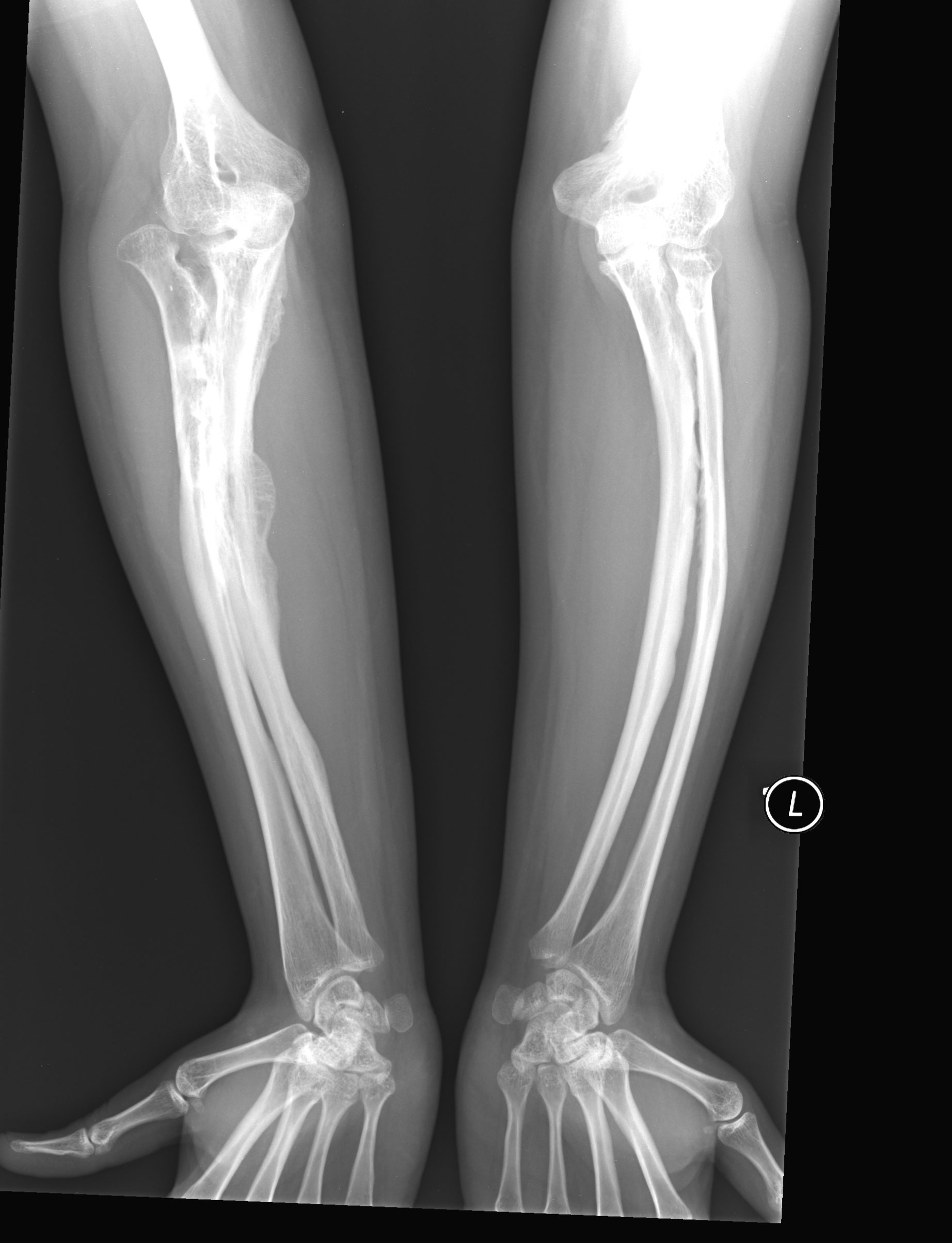
osteogenesis imperfecta type v

Synonyms of Osteogenesis Imperfecta

  • Brittle Bone Disease
  • Fragilitas Ossium
  • Osteopsathyrosis
  • Lobstein’s Disease

Introduction

Osteogenesis Imperfecta is a heterogenous group of disorder with Autosomal Dominant Inheritance Pattern

Etiology of Osteogenesis Imperfecta

etiology of osteogenesis imperfecta

Classification of Osteogenesis Imperfecta

Given by Sillence

Type 1

  • Most common and mildest
  • 10% Prenatal Fracture
  • Blue sclera, Hearing loss
  • Fragile bone, Kyphoscoliosis
  • Sometimes opalscent dentin

Type 2

  • Most severe
  • Extreme bone fragility and fracture
  • 90% Prenatal fracture

Type 3

  • 50% Prenatal fracture
  • All features of type1 except no hearing loss
  • With Opaescent dentin

Type 4

It has 2 subtypes

  • Opalescent dentin is absent
  • Opalescent dentin is present

Clinical Feature of Osteogenesis Imperfecta

  1. Age of onset - Stillborn
  2. Site - Collagenous body part (ex.Bone, Sclera, Fascia, Tendon, Dentin, Skin Fragile and Porous bone) which are more prone to fracture
  3. Fracture heals by Callus formation
  4. Sclera - Pale blue, thin, pigmented choroid giving blue hue
  5. Hearing loss (Hypoacusis) due to compression of auditory nerve by Osteosclerosis
  6. Hypermobility of Joints due to laxity of ligaments
  7. Bleeding capillaries rare
blue sclerae of patient with osteogenesis

Oral Manifestation of Osteogenesis Imperfecta

  1. Abnormal mesodermal calcified tissue
  2. Frontal and Temporal bossing
  3. Abnormal Dentin formation
  4. Tendency of Class 3 malocclusion, Anterior/Posterior Crossbite and Openbite due to Maxillary hypoplasia
  5. Impaction and Ectopic teeth eruption

Radiographic Feature of Osteogenesis Imperfecta

  1. Bowing / Angulate / Deformed fractured bones
  2. Wormian bone in Skull (multiple small sutural bones fail to fuse)
  3. Mixed bony lesions on OPG
  4. Premature pulp obliteration on IOPA

Histopathologic Feature of Osteogenesis Imperfecta

  1. Retarded or abnormal osteoblatic activity
  2. Abnormal Collagen synthesis in organic matrix
  3. Thin cortex
  4. Immature spongy bone
  5. Delicate trabeculae of cancellous bone with microfractures
  6. Defective microvascular system

Treatment of Osteogenesis Imperfecta

  1. Preventive measures
    • Prenatal Ultrasonography
    • Chorion Villus Prenatal Biopsy
  2. Genetic counselling

Malignant Melanoma

Malignant Melanoma

What is Malignant Melanoma?

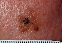
malignant melanoma in situ

Introduction

It is a Malignant Neoplasm of Epidermal Melanocytes and known as most deadliest of Human Neoplasm.

Etiology of Malignant Melanoma

  1. Sunlight Exposure (UV Light)
  2. Artificial UV Source (Tanning Lamp)
  3. Familial - 1st degree relative (2% - 5%)
  4. Xeroderma Pigmentosum
  5. Higher Socioeconomic Status (Outdoor Activities)

Clinicopathologic Classification of Malignant Melanoma

  1. Cutaneous (Most Common)
  2. Non-Cutaneous
  3. Superficial Spreading
  4. Lentigo Maligna
  5. Acral Lentiginous (Most Aggresive)
  6. Amelanotic
  7. Nodular

Clinical Feature of Malignant Melanoma

  1. Incidence - 1/lac/year (Dark Skinned), 50/lac/year (Light Skinned)
  2. Location - Palm, Soles, Mucous Membrane
  3. Precursor Lesion - Acquired Naevus, Dysplastic Naevus, Congenital Naevus, Cellular Blue Naevus

Oral Manifestation Malignant Melanoma

  1. Uncommon in Oral Mucosa (1.6%)
  2. Female Predominance (2×F ˃ M)
  3. Age - 40 - 70 years
  4. Site Predilection - Palate, Maxillary Gingiva, Alveolar Gingiva
  5. Other Sites - Buccal Mucosa, Mandibular Gingiva, Tongue, Lips, Floor of Mouth
  6. Lesion Appearance - Deeply Pigmented, Ulcerated or Hemorrhagic which tends to increase in size
  7. Prodromal Symptoms - Focal Pigmentation (Months or Years back)

Clinical Diagnosis of Malignant Melanoma

( ABCDE Rule )

  • Asymmetry
  • Border - Irregular / Blurred / Notched / Ragged Edges
  • Colour - Pigmentation (Brown / Black /Red / White)
  • Diameter - ˃ 6 mm
  • Elevation - Raised Surface

Histopathologic Feature of Malignant Melanoma

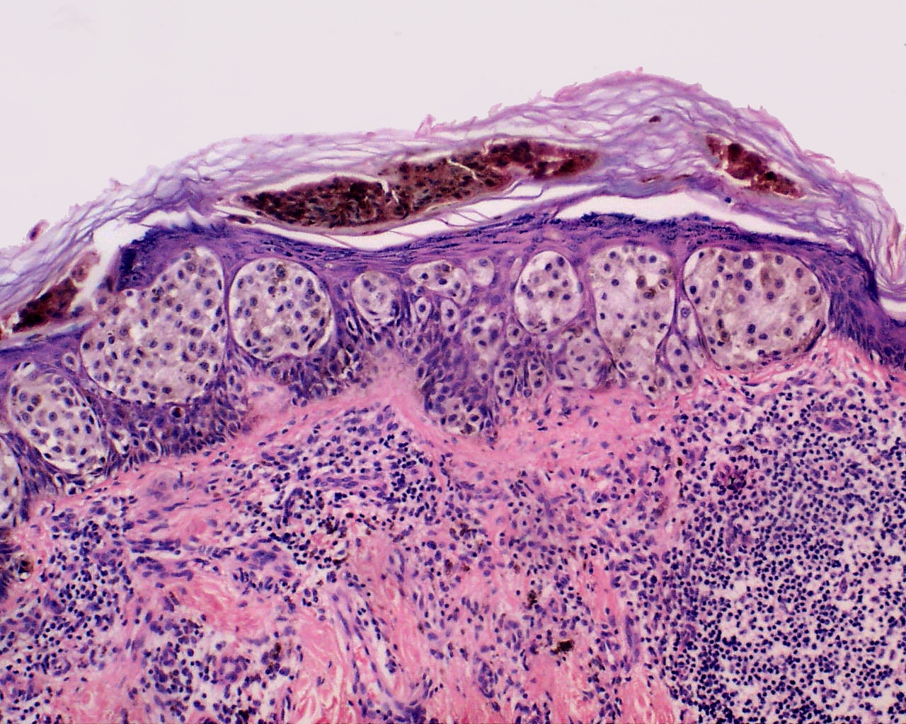
Malignant Melanoma shows Tumor Invasion in deeper histologic cutaneous structure

skin tumors

Clark System (Grades of Tumor Invasion)

Clark Level Characteristics
Level 1 Confined to Epidermis (in situ)
Level 2 Invasion of Papillary Dermis
Level 3 Filling of Papillary Dermis but not extending to Reticular Dermis
Level 4 Invasion of Reticular Dermis
Level 5 Invasion of deep Subcutaneous Tissue

Treatment of Malignant Melanoma

  1. Chemotherapy
  2. Immunotherapy
  3. Surgical - Skin Grafting, Wide Local Incision, Mohs Surgery

Prognosis of Malignant Melanoma

  • Better - Lentigo Maligna (Female Patient)
  • Poor - Nodular, Superficial spreading
  • Worst - Oral Melanotic Lesion

Tracheostomy

Tracheostomy

What is Tracheostomy?

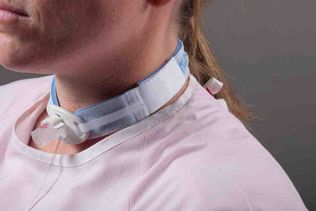
tracheostomy tube

Introduction

Tracheostomy is an operation by which stoma or window is made in the tracheal wall for the purpose of respiration.

Indication of Tracheostomy

  1. Acute and Chronic upper airway obstruction
  2. Maintain patent airway in head and neck surgery
  3. Reduce the risk of pulmonary aspiration
  4. Emergency airway access
  5. Respiratory Paralysis where prolonged intubation is required
  6. Acute Laryngeal Edema eg. Diptheria, Chemical burn and Inhalation of Irritant gases
  7. Excessive tracheobronchial secretion
  8. Foreign body in airway
  9. Injury or pressure to Larynx
  10. Bilateral Abductor Paralysis of vocal cord
  11. Tetanus
  12. Respiratory insufficiency due to Emphysema, Bronchitis, Bronchiectasis

Contraindication of Tracheostomy

  1. Asthma, Chronic Obstructive Pulmonary Disease
  2. Unstable Cervical Spine
  3. Unstable Cardiac Disease
  4. Inability to Cooperate
  5. Severe Hypoxemia

Procedure for Tracheostomy

  1. Preanesthetic medication - Injection atropine 0.6mg half an hour before tracheostomy
  2. Position - The patient is put supine with neck over-extended and chin in the midline
  3. Site - Below isthmus of thyroid gland
  4. Local anesthesia - The part selected is prepared and infiltrated with 2% lignocaine
  5. Procedure - A vertical midline incision is taken about 4 cm long, starting from the suprasternal notch. The skin, platysma and the superficial fascia are cut
  6. The inner margin of sternohyoid muscle is identified and deep layer of cervical fascia is cut
  7. Pretracheal fascia is cut and separated from the trachea
  8. The isthmus is cut between the clamps and its ends ligated
  9. 0.5 ml to 1 ml of lignocaine is injected into the lumen of trachea and fourth and fifth tracheal ring is incised from below upwards
  10. Tracheostomy tube is inserted and tape is tied
  11. The wound of tracheostomy are kept free of secretions by repeated suctions.

Types of Tracheostomy

  1. Emergency
  2. Elective
  3. Permanent

Types of Tracheostomy Tubes

1. Metal Tube : Used for permanent tracheostomy which has inner tube.

metal tracheostomy tube

2. Portex Tube : It has a cuff which can be inflated by injecting the air through outer tube. Balloon gets inflated and secures tracheostomy tube in position.

portex tracheostomy tube

Complications of Tracheostomy

A) During Surgery

  • Injury to trachea and esophagus
  • Injury to recurrent laryngeal nerve
  • False passage

B) Post-operative

  • Blocked tracheostomy tube
  • Surgical emphysema
  • Infection, hemorrhage and ulcerations
  • Pulmonary bronchopneumonia
  • Tracheal stenosis

Cracked Tooth Syndrome

Cracked Tooth Syndrome

What is Cracked Tooth Syndrome?

cracked tooth syndrome

Synonyms for Cracked Tooth Syndrome

  • Cracked Cusp Syndrome
  • Split Teeth Syndrome
  • Incomplete Fracture of Posterior Teeth
  • Greenstick Fracture of Crown

Introduction

A tooth that is incompletely cracked but no part of tooth has broken off.

Pattern of Crack

  1. Centrally located crack may extend to pulp
  2. Peripherally located crack (Cuspal Fracture)

Types of Cracked Tooth Syndrome

  1. Fractured Cusp
  2. Treatable
  3. Non-Treatable
  4. Split Tooth
types of cracked tooth

Clinical Features of Cracked Tooth Syndrome

  1. Pain on biting
  2. Relief after removing pressure
  3. Tenderness positive on Axial Percussion

Mechanism of Pain in Cracked Tooth Syndrome

mechanism of pain in cracked tooth syndrome

Diagnosis for Cracked Tooth Syndrome

  1. Clinical Symptoms
  2. Bite Test - Patient is asked to bite upon Tooth Sloth / Toothpick / Cotton Roll
    Inference - When patient bites on tooth sloth it Pains and which gets relieved when biting pressure has withdrawn.

Risk Factor for Cracked Tooth Syndrome

  1. Heavy Restoration
  2. Extensive Dental Restoration
  3. Occlusal Discrepancies (High Points)
  4. Masticatory Accidents

Treatment for Cracked Tooth Syndrome

  1. Small Crack : Remove Compromised part followed by Restoration
  2. Large Crack (No pulp involvement) : Stabilization with Orthodontic Band / Crown / Cast metal Restoration
  3. Large Crack (With pulp involvement) : Stabilization, Endodontic treatment followed by Permanent Restoration
  4. Split Tooth : Extraction (Hopeless Prognosis)