Obesity | Overweight

Obesity | Overweight

All about Obesity

obesity and overweight

Obesity Introduction

Obesity is widely regarded as a pandemic, with potentially disastrous complication for human health

Causes of Obesity

  1. Increase Energy Intake
    • Increase protein size
    • Increase snacking
    • Loss of regular meals
    • Increase energy food (mainly fat)
    • Increased affluence
  2. Decreasing Energy Expenditure
    • Increase car ownership
    • Decrease walking
    • Increase automation
    • Decrease manual labour
    • Decrease sports in school
    • Increase time spent on computer
    • Increase central heating
  3. Endocrine Factors
    • Hypothyroidism
    • Cushing's Syndrome
    • Insulinoma
    • Hypothalamic tumors / injury
  4. Medications
    • Atypical antipsychotics (eg. Olanzapine)
    • Sulphonylurease
    • Thiazolidinediones
    • Insulin
    • Corticosteroids
    • Sodium valproate
    • Beta blockers

Complications of Obesity

  1. Associated with Diabetes and Hypertension
    • Coronary heart disease
    • Stroke
    • Diabetes complications
  2. Associated with Liver Fat Accumulation
    • Non-alcoholic steatohepatitis
    • Cirrhosis
  3. Associated with Restricted Ventilation
    • Exertional dyspnoea
    • Obstructive sleep apnoea
    • Obesity hypoventilation syndrome
  4. Associated with Mechanical Effects of Weight
    • Urinary incontinence
    • Osteoarthritis
    • Varicose veins
  5. Associated with Increased Peripheral Steroid Interconversion in Adipose Tissue
    • Hormone dependent cancers (breast, uterus)
    • Polycyclic ovarian syndrome
    • Infertility
    • Hirsutism
  6. Others
    • Psychological morbidity (low self esteem, depression)
    • Socioeconomic disadvantages (low income)
    • Gallstones
    • Colorectal cancer
    • Skin infections (groin and submammary candidiasis)

Management of Obesity or Overweight

  1. Lifestyle Advice
    • Regular eating pattern
    • Maximising physical activity
    • More walking
    • Swimming
    • Avoidance of snacking
    • Regular meals
    • Substitution of sweets with artificial sweeteners
  2. Weight loss Diet
    • Low calorie diet
    • Vitamin supplementation
  3. Drugs
    • Orlistat
  4. Surgery
    • Bariatric Surgery
    • Cosmetic Surgical Procedure
    • Apronectomy: Removal of overhanging abdominal skin
  5. Treatment of additional Risk Factors: eg. smoking, excess alcohol consumption, diabetes mellitus, hypertension, obstructive sleep apnoea, hyperlipidaemia

Diabetic Ketoacidosis (DKA)

Diabetic Ketoacidosis

What is Diabetic Ketoacidosis?

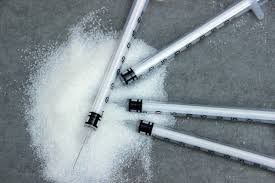
sugar and insulin syringe for diabetes

Introduction

Diabetic Ketoacidosis is a medical emergency and remains a serious cause of morbidity, principally in people with type 1 diabetes

Risk Factors for Diabetic Ketoacidosis

  1. Type 1 Diabetes
  2. Cerebral oedema in children and adolescents
  3. Hypokalaemia
  4. Acute respiratory distress syndrome
  5. Acute myocardial infarction
  6. Sepsis
  7. Pneumonia

Pathogenesis of Diabetic Ketoacidosis

  1. Cardinal Biomechanical features
    • Hyperketonemia (>3 mmol/L)
    • Ketonnuria (>2 on standard urine sticks)
    • Hyperglycaemia (blood glucose >11 mmol/L or >200mg/dL)
    • Metabolic acidosis (venous bicarboate <15mmol/L and Venous pH <7.3)
  2. Hyperglycaemia causes profound osmotic diuresis leading to dehydration and electrolyte loss, particularly Potassium and Sodium
  3. Potassium loss is exacerbated by secondary hyperaldosteronism due to reduced renal perfusion
  4. Ketosis result from insulin deficiency exacerbated by elevated catecholamines and other stress hormones
  5. Due to more unmetabolized acidic ketones, it starts accumulating in blood
  6. Metabolic acidosis forces hydrogen ions into cells, displacing potassium ions

Clinical Feature of Diabetic Ketoacidosis

Symptoms of Diabetic Ketoacidosis

  1. Polyuria, Thirst
  2. Weight loss
  3. Weakness
  4. Nausea, Vomitting
  5. Leg cramps
  6. Blurred vision
  7. Abdominal pain

Signs of Diabetic Ketoacidosis

  1. Dehydration
  2. Hypotension (Postural or Supine)
  3. Cold extremities / Peripheral cyanosis
  4. Tachycardia
  5. Air hunger (Kussmaul breathing)
  6. Smell of Acetone
  7. Hypothermia
  8. Confusion / Drowsiness / Coma

Investigations for Diabetic Ketoacidosis

  1. Venous blood: Urea, Electrolytes, Bicarbonates
  2. Urine or Blood analysis for ketones
  3. ECG
  4. Infection screen
    • Full blood count
    • Blood and Urine culture
    • C-reactive protein
    • Chest X-ray

Treatment for Diabetic Ketoacidosis

  1. Insulin
    • A fixed rate IV infusion of 0.1U/kg body weight/hr is recommended
    • If IV not possible soluble insulin can be given by intramuscular injection with loading dose of 10-20 U, followed by 5 U hourly
    • Alternatively fast acting insulin analogue can be given hourly by subcutaneous injection with initial dose of 0.3 U/kg body weight, followed by 0.1 U/kg hourly
  2. Fluid Replacement
    • Rapid fluid replacement for first few hours is recommennded for adults
    • Caution is recommended in children and adolescent due to chances of developing cerebral oedema
  3. Potassium
    • Careful monitoring of potassium is essential to prevent hyperkalaemia or hypokalaemia
    • Potassium replacement usually not recommended in initial treatment because pre-renal failure maybe present secondary to dehydration
    • Cardiac rhythm should be monitored in severe DKA due to risk of electrolyte induced cardiac arrhythmia
  4. Bicarbonate: Adequate fluid and insulin replacement should resolve acidosis

Epilepsy | Seizure

Epilepsy and Seizure

What is Epilepsy?

epilepsy seizure stroke

Introduction

Epilepsy is the tendency to have unprovoked seizures

Definition of Seizure

Seizure can be defined as the occurrence of signs and symptoms due to abnormal, excessive or synchronous neuronal activity in the brain

Triggering Factors of Seizures

  1. Sleep deprivation
  2. Missed dose of anti-epileptic drugs
  3. Alcohol withdrawal syndromes
  4. Recreational drug misuse
  5. Physical and mental exhaustion
  6. Flickering lights (TV and Computer screens)
  7. Intercurrent infections and metabolic disturbance
  8. Loud music, noise, hot baths

Classification of Seizures

  1. Generalized Seizure
    • Tonic-Clonic Seizure
    • Absence Seizures - Typical / Atypical / Absence with special features
    • Myoclonic - Myoclonic / Myoclonic atonic / Myoclonic tonic
    • Clonic
    • Tonic
    • Atonic
    • Myeloid Absence Seizures
    • Eyelid Myoclonia
  2. Focal Seizures
    • Without impairment of consciousness or awareness
    • With impairment of consciousness or awareness
    • Evolving to a bilateral, convulsive seizure - Tonic / Clonic / Tonic-Clonic
  3. Unknown Seizure
    • Epileptic spasm

Causes of Focal Seizures

A) Idiopathic

  1. Benign Rolandic Epilepsy of childhood
  2. Benign Occipital Epilepsy of childhood

B) Focal Structural Lesions

  1. Genetic
    • Tuberous Sclerosis
    • Autosomal dominant frontal lobe epilepsy
    • Autosomal dominant partial epilepsy with auditory features
    • Von Hippel-Lindau disease
    • Neurofibromatosis
    • Cerebral migration abnormalities
  2. Infantile Hemiplegia
  3. Dysembryonic
    • Cortical dysgenesis
    • Sturge-Weber Syndrome
  4. Mesial Temporal sclerosis
  5. Cerebrovascular disease
    • Intracerebral haemorrhage
    • Cerebral infarction
    • Arteriovenous Malformation
    • Cavernous Haemangioma
  6. Tumours (Primary and Secondary)
  7. Trauma
  8. Infective
    • Cerebral abscess
    • Toxoplasmosis
    • Cysticercosis
    • Tuberculoma
    • Subdural empyema
    • Encephalitis
    • HIV
  9. Inflammatory
    • Sarcoidosis
    • Vasculitis

Causes of Generalized Tonic-Clonic Seizures

  1. Generalisation from Focal Seizure
  2. Genetic
    • Inborn errors of metabolism
    • Storage diseases
    • Phakomatosis
  3. Cerebral birth injury
  4. Hydrocephalus
  5. Cerebral anoxia
  6. Drugs
    • Antibiotics: Penicillin, Isoniazid, Metronidazole
    • Antimalarials: Chloroquine, Mefloquine
    • Ciclosporin
    • Cardiac Anti-arrhythmics: Lidocaine, Disopyramide
    • Psychotropic agents: Phenothiazines, Tricyclic anti-depressants, Lithium
    • Amphetamines withdrawal
  7. Alcohol
  8. Toxins
    • Organophosphate
    • Heavy metals (lead, tin)
  9. Metabolic disease
    • Hypocalcaemia
    • Hypoglycaemia
    • Hypomagnesaemia
    • Liver failure
    • Renal failure
  10. Infective
    • Post-infectious encephalopathy
    • Meningitis
  11. Inflammatory
    • Multiple sclerosis
    • SLE
  12. Diffuse degenerative disease
  13. Alzheimer's Disease
  14. Creutzfeldt-Jakob disease

Investigations of Epilepsy

  1. ECG
    • Standard ECG
    • Sleep ECG
    • ECG with special electrodes
  2. CT
  3. MRI
  4. For Metabolic disorder
    • Urea and electrocytes
    • Blood Glucose
    • Serum calcium, Magnesium
  5. For Inflammatory or Infective disorder
    • Full Blood count
    • Erythrocyte Sedimentation Rate
    • C-reactive protein
    • Chest X-ray
    • Serology for syphilis, HIV, Collagen disease
  6. CSF Examination
  7. Ambulatory ECG
  8. Videotelemetry

Management of Epilepsy

  1. First aid Management for Seizure
    • Move person away from danger (fire, water, machinery, furniture)
    • After convulsion ceases turn person into recovery position (semi-prone)
    • Ensure airway is clear or insert anything in mouth
    • If convulsions continue for more than 5 mins or recur without person regaining consciousness, summons urgent medical attention
    • Do not leave the person alone until fully recovered
  2. Immediate Care
  3. Lifestyle Advice
    • Avoid triggering factors
    • Only shallow bath
    • Prolong cyclone journey is avoided
    • Activities requiring prolonged proximity to water (swimming, fishing, boating) should be carried out in company with someone
  4. Anticonvulsant / Anti-epileptic Therapy
  5. Monitoring Therapy
  6. Epilepsy Thearpy
  7. Withdrawal of Anticolvusant Therapy

Raynaud's Disease / Raynaud's Phenomenon

Raynauds's Disease / Raynaud's Phenomenon

What is Raynaud's Disease?

Raynaud's Syndrome

Introduction

Raynaud's Disease is a type of arterial disease also known as Raynaud's Phenomenon and Raynaud's Syndrome

Definition

Raynaud's Disease is a condition characterized by episodic attacks of vasospasm that causes closure of small arteries and arterioles of distal parts of the extremities in response to cold exposure or emotional stimuli

Etiology and Secondary causes of Raynaud's Disease

  1. Cold climate is precipitating factor
  2. Atherosclerosis
  3. Scleroderma
  4. Systemic lupus
  5. Cervical rib
  6. Carpal tunnel syndrome
  7. Vibrating tools
  8. Thoracic outlet syndrome
  9. Cryoglobulinemia
  10. Dermatomyositis

Pathophysiology of Raynaud's Disease

  1. Stage of Syncope: Arterioles undergoes constriction as an abnormal response to cold, hence affected limb becomes blanched and severe pallor develops
  2. Stage of Asphyxia: After a brief period of vasoconstriction, capillaries dilate, filling with deoxygenated blood resulting in bluish discoloration (Cyanosis) of affected part
  3. Stage of Recovery: After vasospasm subsides, vasodilation occurs with reactive hyperemia which produces redness (rubor)

Clinical Features of Raynaud's Disease

  1. Raynaud's Disease affects young females
  2. It causes bilateral episodic digital ischaemia on exposure to cold
  3. Thumb is usually spared
  4. Peripheral pulses are normal
  5. Pallor, Cyanosis and rubor shows colour changes during attack
  6. Sometimes gangrenous patches seen on fingertips due to recurrent attack

Investigation of Raynaud's Disease

  1. Digital Plethysmography
  2. Arteriography
  3. Haemogram
  4. Protein Electrophoresis

Treatment of Raynaud's Disease

A) Conservative Treatment

  • Moving to warm climate
  • Stoppage of Tobacco
  • Avoid unnecessary exposure to cold
  • Occupational exposure should be reduced
  • Calcium antagonist such as Nifedipine

B) Surgical Treatment

  • Cervico-Thoracic Sympathectomy
  • Cervico-Dorsal Sympathectomy
  • Removal of all rami communicating to 2nd or 3rd thoracic ganglion

Biotransformation | Drug Metabolism

Biotransformation | Drug Metabolism

What is Biotransformation?

drug molecules

Introduction

Biotransformation is chemical alteration of the drug in the body

Steps in Biotransformation Reaction

  1. Inactivation
    • More drugs and there active metabolites are rendered inactive or less active
  2. Active metabolite from inactive drug
    • Many drugs are found to be partially converted to one or more active metabolites
    • The effects observed are the sum total of that due to the parental drug and its active metabolites
  3. Activation of inactive drug
    • Few drugs are inactive and requires conversion in the body to one or more active metabolite
    • These drugs are called Prodrugs, which offers advantage over the active form in being more stable, having better bioavailability or other desirable pharmacokinetic properties
    • Prodrugs show less side effects and toxicity

Classification of Biotransformation Reaction

  • Nonsynthetic Reaction
    1. Oxidation
      • It involves addition of oxygen (negatively charged radical) or removal of hydrogen (positively charged radical)
      • It is most important drug metabolizing reaction
      • It is carried out by a group of monooxygenase in liver which are Cytochrome P-450 haemoprotein, NADPH, cytochrome P-450 reductase and molecular O2
      • eg. Barbiturates, Paracetamol, Phenothiazines, Steroids, Adrenaline
    2. Reduction
      • It is converse of oxidation reaction
      • It involves cytochrome P-450 in opposite direction
      • eg. Chloramphenicol, Halothane, Warfarin
    3. Hydrolysis
      • It shows breakdown of drug molecule by taking up a water molecule
      • Sites for hydrolysis are liver, intestine, plasma
      • eg. Choline esters, Procaine, Lidocaine, Oxytocin
  • Synthetic Reaction
    1. Glucuronide Conjugation
      • It is carried out by a group of UDP-glucuronosyl transferase (UGTs)
      • Compounds with hydroxyl or carboxylic acid are easily conjugated with glucuronic acid
      • eg. Chloramphenicol, Aspirin, Morphine, Metronidazole
    2. Acetylation
      • Compounds having amino or hydralazine residue are conjugated with the help of acetyl coenzyme-A
      • eg. Sulfonamides, Isoniazid, Hydralazine
    3. Methylation
      • Amines and phenols can be methylated
      • Methionine and cysteine acts as methyl donors
      • eg. Adrenaline, Histamine, Nicotinic acid
    4. Sulfate Conjugation
      • Phenolic compound drugs are sulfated by sulfotransferase
      • eg. Chloramphenicol, Steroids
    5. Glycine Conjugation
      • Drugs having carboxylic acids are conjugated with glycine
      • eg. Salicylates
    6. Glutathione Conjugation
      • It serves to deactivate highly reactive quinone or epoxide intermediates formed during metabolism of certain drugs
      • eg. Paracetamol
    7. Ribonucleoside / Nucleotide Synthesis
      • It is important for activation of many purine and pyrimidine antimetabolites used in cancer chemotherapy

Types of Drug Metabolizing Enzyme

  1. Microsomal
    • These are located on smooth endoplasmic reticulum eg. Liver, Kidney, Intestinal mucosa, Lungs
    • eg. Monooxygenase, Cytochrome P-450, UGTs
    • They catalyst most of the oxidation, reduction, hydrolysis, and glucuronide conjugation
  2. Nonmicrosomal
    • These are located in cytoplasm, mitochondria of hepatic cells
    • eg. flavoprotein oxidase, esterase, amidase and conjugase
    • They catalyst oxidation, reduction and hydrolytic reactions

Hoffman Elimination

It refers to inactivation of the drug in the body fluids by spontaneous molecular rearrangement without the agency of any enzyme eg. atracurium

Diabetes Mellitus

Diabetes Mellitus

All about Diabetes Mellitus

diabetes checking machine

Introduction

Diabetes Mellitus is a clinical syndrome characterized by an increase in plasma blood glucose level, which is also called as Hyperglycemia

Etiological Classification of Diabetes Mellitus

  1. Type 1 Diabetes (Insulin Dependent Diabetes Meliitus)
    • Immune mediated
    • Idiopathic
  2. Type 2 Diabetes (Insulin Independent Diabetes Mellitus)
  3. Other Specific Types
    • Genetic defect of beta cell function
    • Genetic defect of insulin action
    • Pancreatic disease
    • Excess endogenous production of hormone antagonist to insulin
    • Drug induced
    • Uncommon form of immune mediated diabetes
    • Associated with genetic syndromes
  4. Gestational Diabetes

Symptoms of Hyperglycemia

  1. Thirst
  2. Dry Mouth
  3. Nocturia
  4. Unusual weight loss
  5. Tiredness, Fatigue, Lethargy
  6. Blurring of vision
  7. Nausea
  8. Headache
  9. Hyperphagia - Predilection for sweet foods
  10. Mood change, irritability
  11. Difficulty in concentrating
  12. Apathy
  13. Genital candidiasis - pruritus vulve, balanitis

Diagnosis of Diabetes and Pre-Diabetes

  1. Plasma Glucose level
  2. Plasma Urea, Creatinine, Electrolytes
  3. Liver Function Test
  4. Thyroid Function Test
  5. Urine test for Ketones, protein or microalbuminuria

Management of Diabetes Mellitus

  1. Dietary and lifestyle modification
    • Regular physical activity
    • Healthy diet
    • Limit the sodium intake (less than 6 gm per day)
    • Reducing alcohol consumption
    • Stoppage of smoking
  2. Oral Anti-diabetic drugs
    • Biguanide - Metformin
    • Sulphonylurease
    • Alpha Glucosidase inhibitor
    • Thiazolidinediones
    • Sodium and Glucose Transporter 2 (SGLT2) inhibitor
  3. Injectable Insulin therapy

Complications of Diabetes Mellitus

A) Microvascular / Neuropathic

  1. Retinopathy - Cataract, Impaired vision
  2. Nephropathy - Renal failure
  3. Peripheral Neuropathy - Sensory loss, Pain, Motor weakness
  4. Autonomic Neuropathy - GIT problems, Postural hypotension
  5. Foot Disease - Ulceration, Arthropathy

B) Macrovascular

  1. Coronary Circulation - Myocardial Ischaemia / Infarction
  2. Cerebral Circulation - Transient Ischaemic Attack, Stroke
  3. Peripheral Circulation - Claudication, Ischaemia

Hypertension

Hypertension

All about Hypertension

blood pressure measurement

Introduction

Hypertension is the level of blood pressure (BP) at which the benefits of treatment outweigh the costs and hazards

Types of Hypertension

Staging of Hypertension Systolic BP Diastolic BP
Prehypertension 120 - 139 mmHg 80 - 89 mmHg
Stage 1 Hypertension 140 - 159 mmHg 90 - 99 mmHg
Stage 2 Hypertension 160 - 179 mmHg 100 - 109 mmHg
Stage 3 Hypertension >180 mmHg >110 mmHg

Causes of Secondary Hypertension

  1. Alcohol
  2. Obesity
  3. Renal Disease
    • Parenchymal Renal Disease - Glomerulonephritis
    • Renal Vascular Disease
    • Polycystic Kidney Disease (PKD)
  4. Endocrine Disease
    • Pheochromocytoma
    • Cushing's Syndrome
    • Thyrotoxicosis
    • Primary Hyperaldosteronism (Conn's Syndrome)
    • Hyperparathyroidism
    • Primary Hyperthyroidism
    • Acromegaly
    • Liddle's Syndrome
    • Congenital Adrenal Hyperplasia (CAH)
  5. Drugs
    • Oral Contraceptives containing oestrogen
    • Anabolic Steroids
    • Corticosteroids
    • NSAIDs
    • Carbenoxolone
    • Sympathomimetic Agent
    • Coarctation of Aorta

Symptoms of Hypertensive Patients

  1. Severe headache
  2. Nosebleed
  3. Fatigue or confusion
  4. Vision problem
  5. Chest pain
  6. Difficulty in breathing
  7. Irregular heartbeat
  8. Blood in the urine
  9. Pounding in your chest, neck or ears
  10. Dizziness
  11. Nervousness
  12. Sweating
  13. Trouble sleeping
  14. Facial flushing
  15. Blood spots in eyes

Signs of Hypertension

  1. Radiofemoral Delay (Coarctation of Aorta)
  2. Enlarged Kidneys (Polycystic Kidney Disease)
  3. Abdominal Bruits (Renal Artery Stenosis)
  4. Characteristic Facies (Cushing's Syndrome)
  5. Blood vesels Atherosclerosis
    • Larger arteries (more than 1 mm in diameter): Internal elastic lamina is thickened, Smooth muscle is hypertrophied and Fibrous tissue is deposited. So vessels dilates and becomes torus with less compliant walls
    • Smaller arteries (less than 1 mm in diameter): Hyaline arteriosclerosis in wall, narrowing of lumen
    • Widespread atheroma develop and may lead to coronary and cerebrovascular diseases
    • These vascular changes increase peripheral vascular resistance which aggravates hypertension. Reduced renal blood flow activates Renin-Angiotensin-Aldosterone-Axis
  6. Central Nervous System
    • Cerebral Haemorrhage and Cerebral Infarction leads to Stroke Carotid Atheroma Subarachnoid Haemorrhage Hypertensive Encephalopathy
  7. Retina
    • Cotton wool exudates and Microaneurysm
    • Retinal ischaemia and infarction
    • Central retinal vein thrombosis
  8. Heart
    • Left ventricular hypertrophy
    • Atrial fibrillation
    • Left ventricular failure
  9. Kidneys
    • Proteinuria
    • Renal failure

Diagnosis of Hypertensive Patients

  1. Accurate BP measurement
  2. Identify contributing factors and underlying causes
  3. Detect any complications eg.target organ damage (blood vessels, CVS, CNS, Retina, Kidney)

Investigations of Hypertensive Patients

  1. Urinalysis of blood, protein and glucose
  2. Blood, urea, electrolytes and creatinine
  3. Blood Glucose
  4. Serum total and HDL Cholesterol
  5. Thyroid Function Tests
  6. 12 - Lead ECG

Management of Hypertensive Patients

  1. Quantification of Cardiovascular Risk
  2. Threshold for Intervention
  3. Treatment Targets
  4. Non-drug Therapy
    • Correcting Obesity
    • Reducing Alcohol intake
    • Quitting Smoking
    • Restricting Salt intake
    • Regular Physical Exercise
    • Increasing consumption
  5. Antihypertensive Drugs
    • Thiazide and other Diuretics
    • ACE Inhibitors
    • Beta blockers
    • Labetalol, Carvedilol
  6. Emergency Treatment for Malignant Hypertension
  7. Adjuvant Drug Therapy
    • Antiplatelet Therapy - Aspirin
    • Statins

Tongue Anatomy

Tongue Anatomy

All about Tongue Anatomy

tongue anatomy and morphology

Introduction

The tongue is a muscular organ situated in the floor of the mouth

Parts of Tongue

  1. Root
    • Root is attached to the styloid process and soft palate above, and to mandible and the hyoid bone (lingual bone or tongue-bone) below
    • Tongue cannot be swallowed due to these attachments, related to geniohyoid and mylohyoid attachments
  2. Tip: Tip of tongue forms the anterior free end, which lies behind the upper incisor teeth at rest position
  3. Dorsum / Body: It is convex in all directions and divided into following 3 parts
    • Oral or Papillary part of the tongue
      • It is the anterior 2/3rd part of tongue
      • It is placed on the floor of the mouth
      • It's margins are free and in contact with gums and teeth
      • In front of palatoglossal arch it's both margins shows 4 to 5 vertical folds named Foliate Papillae
      • Superior surface shows a median furrow and covered with papillae which makes it rough
      • Inferior surface shows a median fold called Frenulum Linguae, covered by a smooth mucous membrane
      • On either side of frenulum there is a prominence produced by the deep lingual vein and laterally there is a fold called Plica Fimbriata
    • Pharyngeal / Lymphoid part of the Tongue
      • It lies behind palatoglossal arch and sulcus terminalis
      • It's posterior part is called base of the tongue, which forms the anterior wall of oropharynx
      • It's mucous membrane has no papillae, but it has many lymphoid follicles that collectively constitutes the lingual tonsil
    • Posterior most part of the Tongue
      • It is connected to the epiglottis by 3 folds of mucous membrane, which are median glossoepiglottic fold and the right and left lateral glossoepiglottic fold
      • On either side of median fold, there is depression called Vallecula
      • Lateral folds separates the vallecula from the piriform fossa

Clinical Anatomy of Tongue

  1. Glossitis: Inflammation of tongue, which is a part of generalized oral cavity ulceration or stomatitis
  2. Atrophy of filliform papillae: Tongue becomes smooth in certain anaemias
  3. Acute Glossitis: Presence of rich lymphatics network and loose areolar tissue is responsible for enormous swelling of the tongue
  4. Undersurface of tongue is good site to diagnose Jaundice
  5. In unconscious patients, tongue may fall back and obstruct the air passage which can be prevented by lying the patient on one side with head down or keeping the tongue out mechanically

Papillae of Tongue

Tongue Papillae are the projections of mucous membrane or corium which gives the roughness to anterior 2/3rd of tongue

  1. Circumvallate / Vallate Papillae
    • Largest size of all papillae, with 1 - 2 mm in diameter
    • 8 - 12 in number
    • Situated immediately anterior to the sulcus terminalis
    • Each papilla is a cylindrical projection surrounded by a circular sulcus
    • The wall of papillae has taste buds
  2. Fungiform Papillae
    • These are numerous at tip and margins of the tongue, sometimes also seen in dorsum of tongue
    • Smaller than circumvallate papillae but larger than filliform papillae
    • Each papilla contains narrow pedicle and a large rounded head
    • They have bright red colour
  3. Filliform / Conical Papillae
    • Covers the presulcular area of dorsum of the tongue, giving it's characteristic Velvety appearance
    • Smallest and most numerous of the lingual papillae
    • Each papillae is pointed and covered with keratin
  4. Folliate Papillae: 4 to 5 vertical folds at lingual margin

Muscles of Tongue

Middle fibrous septum divides the tongue into left and right halves, each half contain 4 Intrinsic and 4 Extrinsic muscles as follows:

  1. Intrinsic Muscles
    • Superior Longitudinal
    • Inferior Longitudinal
    • Transverse
    • Vertical
  2. Extrinsic Muscles
    • Genioglossus
    • Hyoglossus (often including the Chondroglossus)
    • Styloglossus
    • Palatoglossus

Arterial Supply of Tongue

  1. It is derived from the tortuous Lingual Artery, branch of the External Carotid Artery
  2. Root of tongue is supplied by Tonsillar Artery, branch of Facial Artery and Ascending Pharyngeal branch of External Carotid Artery

Venous Drainage of Tongue

  1. Deep lingual vein is largest and main vein of tongue
  2. It unites the Lingual vein which ends in the Internal Jugular Vein

Lymphatic Drainage of Tongue

  1. Tip of tongue bilaterally drains to Submental lymph node
  2. Anterior 2/3rd of tongue drains to Submandibular lymph node
  3. Posterior 1/3rd and posterior most part drains to upper deep cervical and jugulodigastric lymph node
  4. Whole lymph finally drains to Jugulo-omohyoid lymph node; known as Lymph Node of Tongue

Nerve Supply of Tongue

  1. Motor Nerve: All the intrinsic and extrinsic muscles are supplied by the Hypoglossal nerve except Palatoglossus which is supplied by Cranial root of the accessory nerve through the Pharyngeal nerve plexus
  2. Sensory Nerve:
    • Anterior 2/3rd of tongue: Lingual nerve for general sensation and Chorda tympani for taste sensation, except circumvallate papillae
    • Posterior 1/3rd of tongue: Glossopharyngeal nerve for both general sensation and for taste sensation, including circumvallate papillae
    • Posteriormost of tongue: Vagus nerve through internal laryngeal branch

Histology of Tongue

  1. Bulk of the tongue is made up of striated muscles
  2. Mucous membrane consist of layer of connective tissue, lined by stratified squamous epithelium. On oral part of dorsum it is thin, forms papillae and adherent to the muscles. On pharyngeal part of dorsum, it is very rich in lymphoid follicles. Numerous mucous and serous gland lies deep to the mucous membrane
  3. Taste buds are most numerous on the sides of circumvallate papillae and walls of surrounding sulci. Taste buds are numerous over the foliate papillae and posterior 1/3rd of tongue, whereas sparsely distributed on the fungiform papillae, soft palate, epiglottis and pharynx. There are no taste buds on mid dorsal region of oral part of tongue

Development of Tongue

  1. Epithelium
    • Anterior 2/3rd: First 2 lingual swellings arises from 1st branchial arch
    • Posterior 1/3rd: 3rd branchial arch
    • Posterior most: 4th branchial arch
  2. Muscle: Occipital myotomes
  3. Connective tissue: Local mesenchyme

Taste Pathway of Tongue

  1. Taste from anterior 2/3rd of tongue except circumvallate papillae is carried by Chorda tympani till the geniculate ganglion. The central process go to the tractus solitarius in the medulla
  2. Taste from posterior 1/3rd of tongue including circumvallate papillae is carried by Glossopharyngeal nerve till the inferior ganglion. The central processes also reach the tractus solitarius
  3. Taste from the posterior most part of the tongue and epiglottis is carried by Vagus nerve till inferior ganglion. These central processes also reach tractus solitarius
  4. After a relay in tractus solitarius, the solitario-thalamic tract is formed which becomes a part of trigeminal lemniscus and reaches postero-ventromedial nucleus of thalamus of the opposite side. Another relay here takes them to lowest part of post central gyrus, which is the area for taste